
IMAGE
Fig. S2
- ID
- ZDB-IMAGE-180827-36
- Publication
- Astone et al., 2018 - Zebrafish mutants and TEAD reporters reveal essential functions for Yap and Taz in posterior cardinal vein development
- All Figures
- Figures for Astone et al., 2018
Image
|
Figure Caption
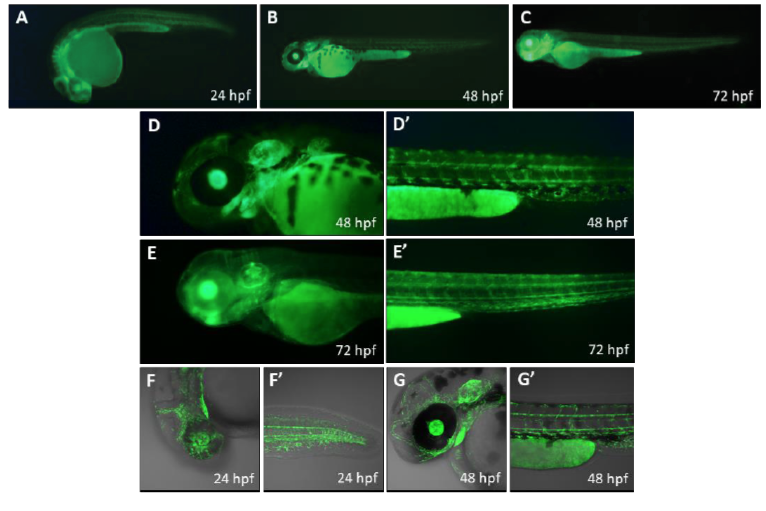
Fig. S2
Overview of the Tg(Hsa.CTGF:eGFP) reporter expression.
In vivo fluorescent microscope (A-E’) and confocal images (F-G’) of Tg(Hsa.CTGF:eGFP) larvae at 24, 48 and 72 hpf.
Acknowledgments
This image is the copyrighted work of the attributed author or publisher, and
ZFIN has permission only to display this image to its users.
Additional permissions should be obtained from the applicable author or publisher of the image.
Full text @ Sci. Rep.