
Image
|
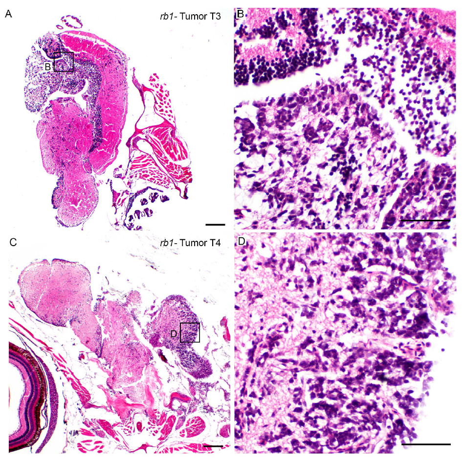
Figure Caption
Fig. S1
Histological analysis of neoplastic populations in TALEN induced rb1-brain tumor T3 and T4 is consistent with primitive neuroectodermal-like tumor. H&E stained sections of tissue remaining after dissection of tumors T3 (A, B) and T4 (C, D) used for transcriptome analysis. Sections exhibit an unencapsulated, multifocally infiltrative neoplastic population within the neuropil. Neoplastic cells are small with deeply basophilic, round to oval to wedge-shaped nuclei, densely clumped chromatin, and scant amounts of cytoplasm. Scale bars A, C 200μm; B, D 50 μm.
Acknowledgments
This image is the copyrighted work of the attributed author or publisher, and
ZFIN has permission only to display this image to its users.
Additional permissions should be obtained from the applicable author or publisher of the image.
Full text @ Dis. Model. Mech.