
Fig. 3
- ID
- ZDB-IMAGE-180724-38
- Publication
- Pogoda et al., 2018 - Direct activation of chordoblasts by retinoic acid is required for segmented centra mineralization during zebrafish spine development
- All Figures
- Figures for Pogoda et al., 2018
|
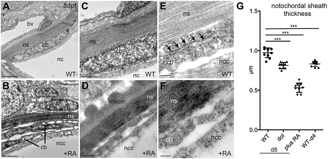
Fig. 3
Chordoblasts of RA-treated zebrafish larvae acquire preosteocyte-like morphological features. (A-F) Transmission electron micrographs of transverse sections through the notochord of control- (A,C,E; level of prospective intervertebral spaces) and RA-treated (B,D,F) larvae at 8 dpf. Scale bars: 2 µm (A,B); 0.5 µm (C,D); 0.1 µm (E,F). Note the higher electron density of the notochord sheath in B and D, indicative of its strong mineralization. bv, blood vessel; cb, chordoblast; mecm, mineralized extracellular matrix; nc, notochord; ncc, notochordal cell; ns, notochordal sheath. Arrows in E point at a stretch of rough endoplasmic reticulum, which is barely found in chordoblasts of RA-treated larvae (F). (G) Quantification of notochord sheath thickness in different genetic backgrounds or after RA treatment as indicated; n=10 per condition; mean±s.d.; ***P<0.0001. dol, cyp26b1ti230g mutant (dolphin) (Laue et al., 2008). At 8 dpf (d8), the mutant shows reduced thickness of the notochord sheath, comparable to the thickness of wild types at 4 dpf (d4), most likely due to the premature reduction of sheath formation in chordoblasts. In wild-type larvae treated with RA from 4 to 8 dpf, the width of the sheath is even more strongly reduced, possibly pointing to additional RA-induced active sheath resorption (compare with Jeradi and Hammerschmidt, 2016; To et al., 2012).