
|
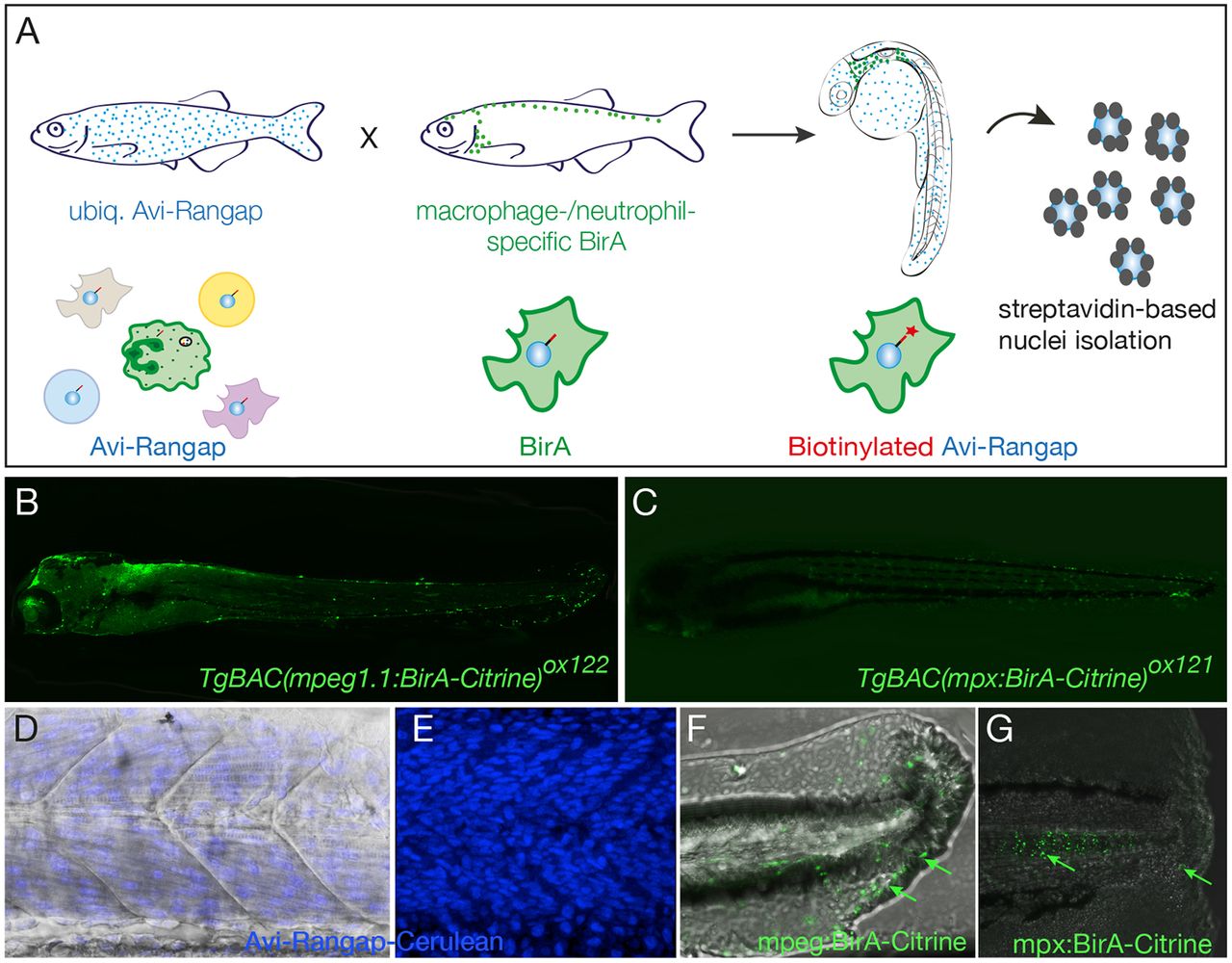
Fig. 3
Binary transgenic zebrafish model for regulatory profiling of myeloid cells. (A) Schematic of myeloid nuclear biotagging system. When biotagging effector transgenic zebrafish line ubiquitously expressing Avi-tagged Rangap for biotinylation of nuclei is crossed to biotagging driver line expressing BirA in myeloid-specific manner, only the macrophage or neutrophil nuclei will be biotinylated. (B) Transgene expression in macrophage-specific BirA driver, TgBAC(mpeg1:BirA-Citrine)ox122, is amplified with anti-GFP–Alexa-Fluor-488 to show expression in macrophages. (C) TgBAC(mpx:BirA-Citrine)ox121 biotagging transgenic driver shows neutrophil-specific expression. (D,E) Projection of confocal microscope images of Tg(bactin:Avi-Cerulean-Rangap)ct700a shows ubiquitous expression of the biotag effector specifically on nuclei, across somite region of the embryo, with (D) and without (E) bright field background. The images in D and E are taken in different embryos. (F,G) Tailfins of transgenic fish transected at 3 dpf show responding macrophages in mpeg1:BirA-Citrine (F) and neutrophils in mpx:BirA-Citrine (G) larvae at 5 hpi or 1 hpi, respectively, as indicated by green arrows.