
|
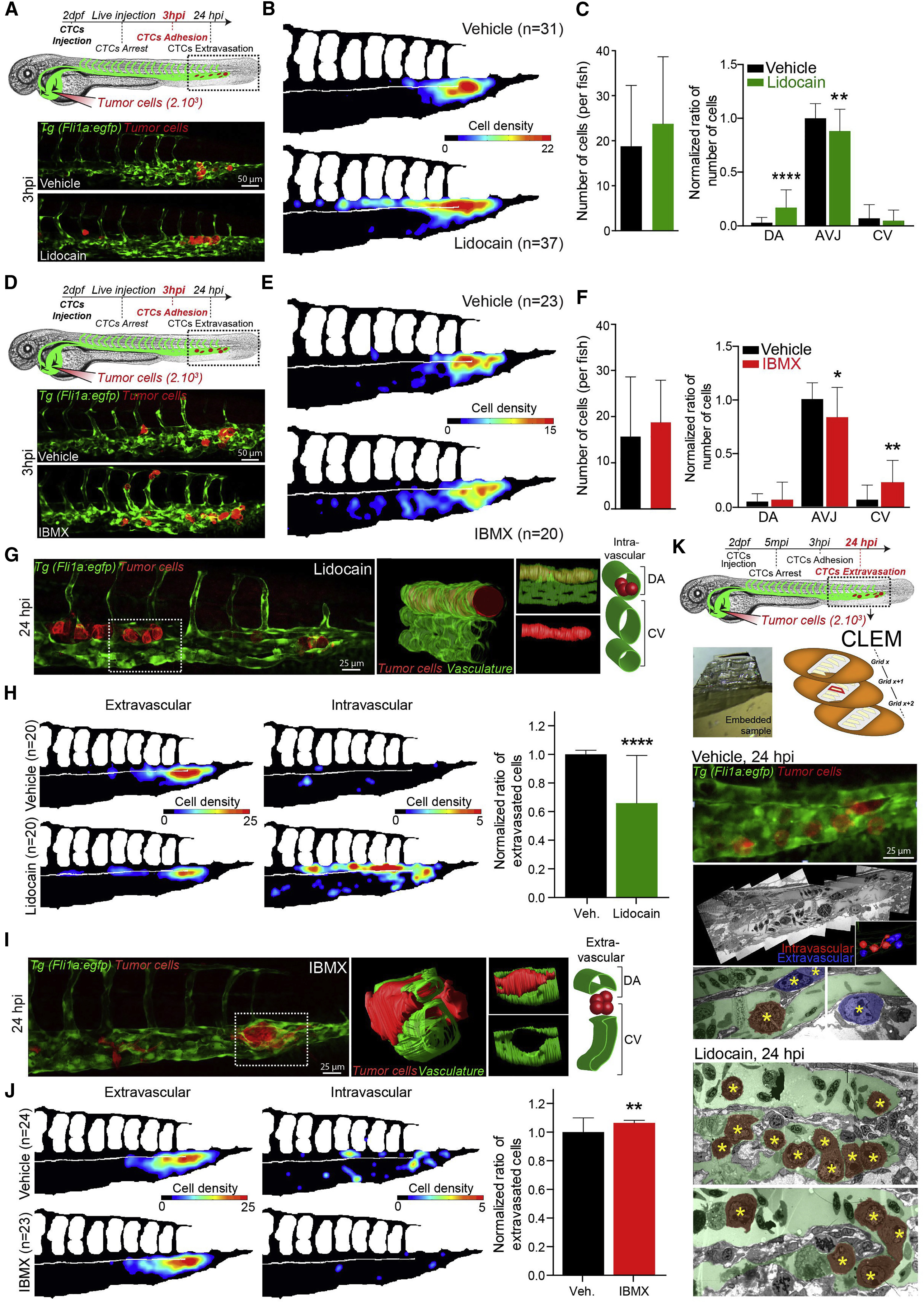
Fig. 4
Modulating Blood Flow Forces Regulates Stable Adhesion and Extravasation of CTCs
(A–J) Zebrafish embryos were treated with vehicle, lidocain (A–C and G–H) or IBMX (D–F and I–J). (A and D) Experimental workflow and representative images of arrested CTCs (red) in the CP (green) of vehicle- or drug-treated embryos. (B and E) Quantification of the number and location of arrested CTCs in the CP of 3 hpi vehicle- or drug-treated embryos, through heatmapping. (C and F) Quantification of the number of arrested CTCs per embryo as well as the ratio of arrested CTCs per region in vehicle- or drug-treated embryos, 3 hpi. Data normalized to vehicle AVJ mean ratio. (G and I) Representative images of TCs (red) in the CP (green) of 24 hpi vehicle- or drug-treated embryos are shown. 3D reconstruction and scheme of the boxed region is provided. (H and J) Quantification of the number and location of intra- and extravascular TCs in the CP of 24 hpi vehicle- or drug-treated embryos, through heatmapping and histograms. (K) Experimental workflow and CLEM analysis of 24 hpi vehicle- or lidocain-treated embryos is used for further assessing the vascular location of TCs (yellow stars) intravascular (red) or extravascular (blue) (see also Video S12).
Values are mean ± SD. ∗p < 0.05, ∗∗p < 0.01, ∗∗∗∗p < 0.0001.
Reprinted from Developmental Cell, 45, Follain, G., Osmani, N., Azevedo, A.S., Allio, G., Mercier, L., Karreman, M.A., Solecki, G., Garcia Leòn, M.J., Lefebvre, O., Fekonja, N., Hille, C., Chabannes, V., Dollé, G., Metivet, T., Hovsepian, F., Prudhomme, C., Pichot, A., Paul, N., Carapito, R., Bahram, S., Ruthensteiner, B., Kemmling, A., Siemonsen, S., Schneider, T., Fiehler, J., Glatzel, M., Winkler, F., Schwab, Y., Pantel, K., Harlepp, S., Goetz, J.G., Hemodynamic Forces Tune the Arrest, Adhesion, and Extravasation of Circulating Tumor Cells, 33-52.e12, Copyright (2018) with permission from Elsevier. Full text @ Dev. Cell