
Fig. S5
- ID
- ZDB-IMAGE-180705-58
- Publication
- Williams et al., 2018 - Gon4l regulates notochord boundary formation and cell polarity underlying axis extension by repressing adhesion genes
- All Figures
- Figures for Williams et al., 2018
|
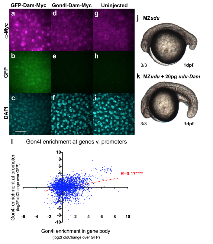
Fig. S5
Dam fusion proteins are functional
a-i) Anti-Myc Immunofluorescent staining for Dam-Myc fusion proteins in WT embryos injected with gfp-dammyc (a-c) or udu-dam-myc (d-f) RNA or uninjected controls (g-i) at blastula stage. Images are representative of multiple embryos from a single trial. j) Uninjected MZudu-/- sibling at 1 dpf. k) MZudu-/- injected with 20pg ududam RNA. Fractions indicate number of embryos with the pictured phenotype over the number of embryos examined. l) Correlation between Gon4l enrichment across promoters and enrichment across gene bodies of genes with regions of significant Gon4l association (Spearmann correlation ****p<0.0001). Dotted line is linear regression of correlation. Negative values indicate depletion of Gon4l relative to GFP controls. Scale bar is 50μm.