
Fig. S3
- ID
- ZDB-IMAGE-180705-56
- Publication
- Williams et al., 2018 - Gon4l regulates notochord boundary formation and cell polarity underlying axis extension by repressing adhesion genes
- All Figures
- Figures for Williams et al., 2018
|
Fig. S3
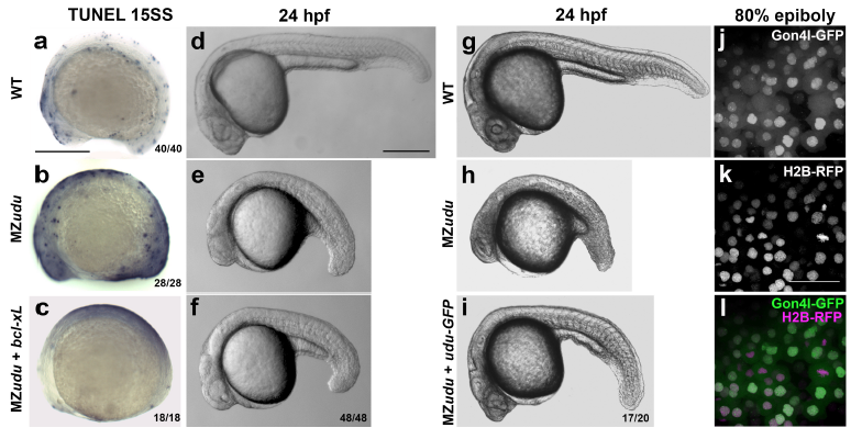
Increased apoptosis in MZudu-/- embryos is not causative of axis extension defects
a-c) TUNEL staining in WT (a), MZudu-/- (b), and MZudu + bcl-xL RNA (c) at 15 somite stage. d-i) Live WT (d,g), MZudu-/- (e,h), MZudu + bcl-xL (f), and MZudu + 25pg udu-gfp (i) embryos at 24hpf. Fractions indicate the number of embryos with the pictured phenotype over the number of embryos examined. j-l) Live WT embryos at 80% epiboly expressing Gon4l-GFP (j) and Histone2B-RFP (k), merged channels in l. Images are representative of four independent trials. Scale bars are 300μm in a-i, 50μm in k. Anterior is to the left.