
IMAGE
Fig. 6
- ID
- ZDB-IMAGE-180702-4
- Genes
- Publication
- Hocking et al., 2018 - Morphogenetic defects underlie Superior Coloboma, a newly identified closure disorder of the dorsal eye
- All Figures
- Figures for Hocking et al., 2018
Image
|
Figure Caption
Fig. 6
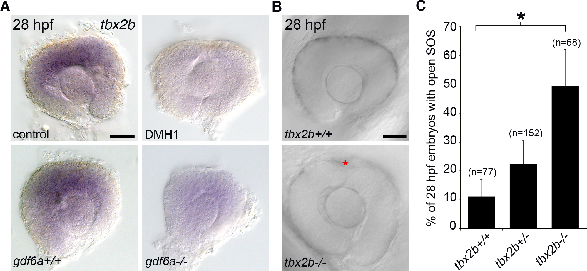
Analysis of Tbx2b and closure of the superior ocular sulcus.
(A) Whole-mount in situ hybridization for zebrafish tbx2b in control and BMP-depleted embryos. Top panels are eyes dissected from control and DMH1-treated embryos; bottom panels are from gdf6a+/+, and gdf6a-/- embryos. (B-C) Analysis of SOS closure in Tbx2b-depleted embryos. DIC images of eyes from live tbx2b+/+ (top panel) and tbx2bfby (bottom panel) embryos (B). Quantification of SOS closure in wild type and tbx2bfby mutant zebrafish eyes (C). Data are means ± SEM; one-way ANOVA with Tukey’s test: *P<0.05. Scale bars are 50 μm.
Figure Data
Acknowledgments
This image is the copyrighted work of the attributed author or publisher, and
ZFIN has permission only to display this image to its users.
Additional permissions should be obtained from the applicable author or publisher of the image.
Full text @ PLoS Genet.