
|
Fig. S3
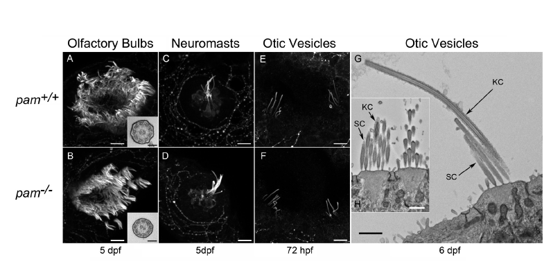
Cilia are present in the olfactory bulbs, neuromasts and otic vesicles of pam-/- embryos
Confocal immunofluorescence images of the olfactory bulb (A, B), neuromast mechano-sensory cells (C, D), and otic vesicles of control and pam-/- embryos probed with antibody against acetylated tubulin. Electron micrographs (G, H) revealed that at 6 dpf both kinocilia (KC) and actin-based stereocilia (SC) were evident emanating from the sensory hair cells in the otic vesicles of pam-/- embryos; these structures assemble early in development at ~16 hpf. The insets in (A, B) show that the olfactory motile cilia have normal 9+2 ultrastructure. Bars = 5 μm (A-F), 1 μm (G, H) and 100 nm (insets in A and B).