
Fig. S3
- ID
- ZDB-IMAGE-180622-36
- Publication
- Missinato et al., 2018 - Dusp6 attenuates Ras/MAPK signaling to limit zebrafish heart regeneration
- All Figures
- Figures for Missinato et al., 2018
|
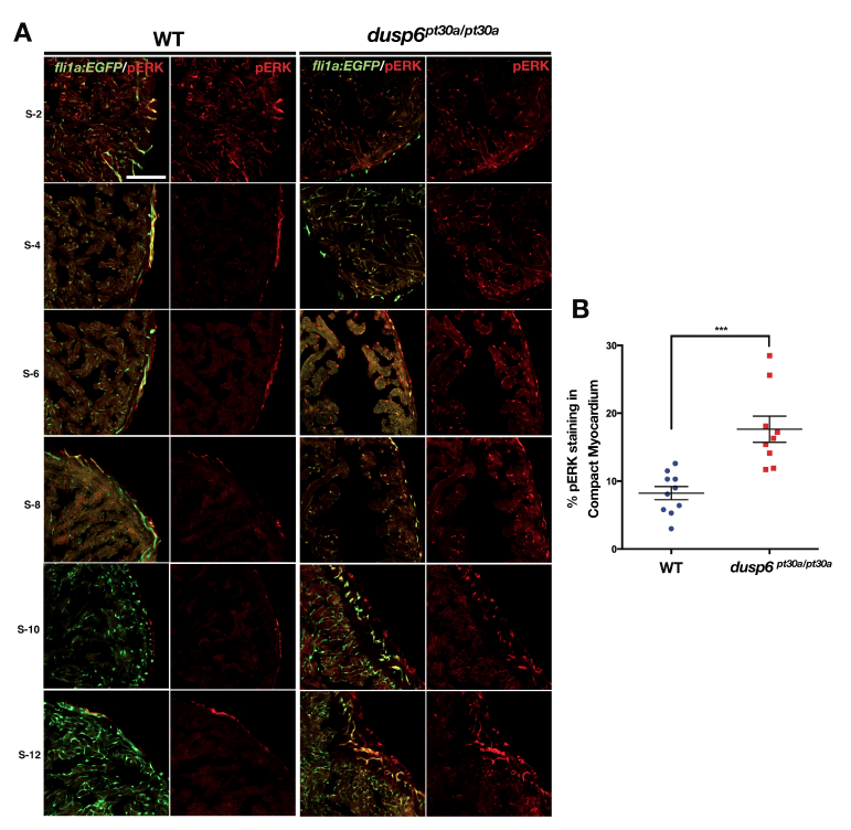
Fig. S3
dusp6 mutant hearts have increased pERK (A) Cryosections of uninjured Tg(fli1a:EGFP)y1 zebrafish hearts immunostained for pERK. dusp6 heart (n=9) have increased pERK staining than WT hearts (n=10). Images are from one representative hearts for each group. Non-consecutive sections (S) are shown. S-2 is an initial section, in proximity of the compact myocardium. S-12 is the central section of the heart. (B) Quantification of pERK in WT and dusp6 mutant hearts at 5 months of age. pERK staining was calculated as sum of pERK+ immunostained area divided by the compact myocardium area, and expressed as percentage. ***p<0.001. Student’s t-test. Scale bars, 100 μm.