
Fig. 5
- ID
- ZDB-IMAGE-180620-20
- Publication
- Auer et al., 2018 - Evidence for Myelin Sheath Remodeling in the CNS Revealed by In Vivo Imaging
- All Figures
- Figures for Auer et al., 2018
|
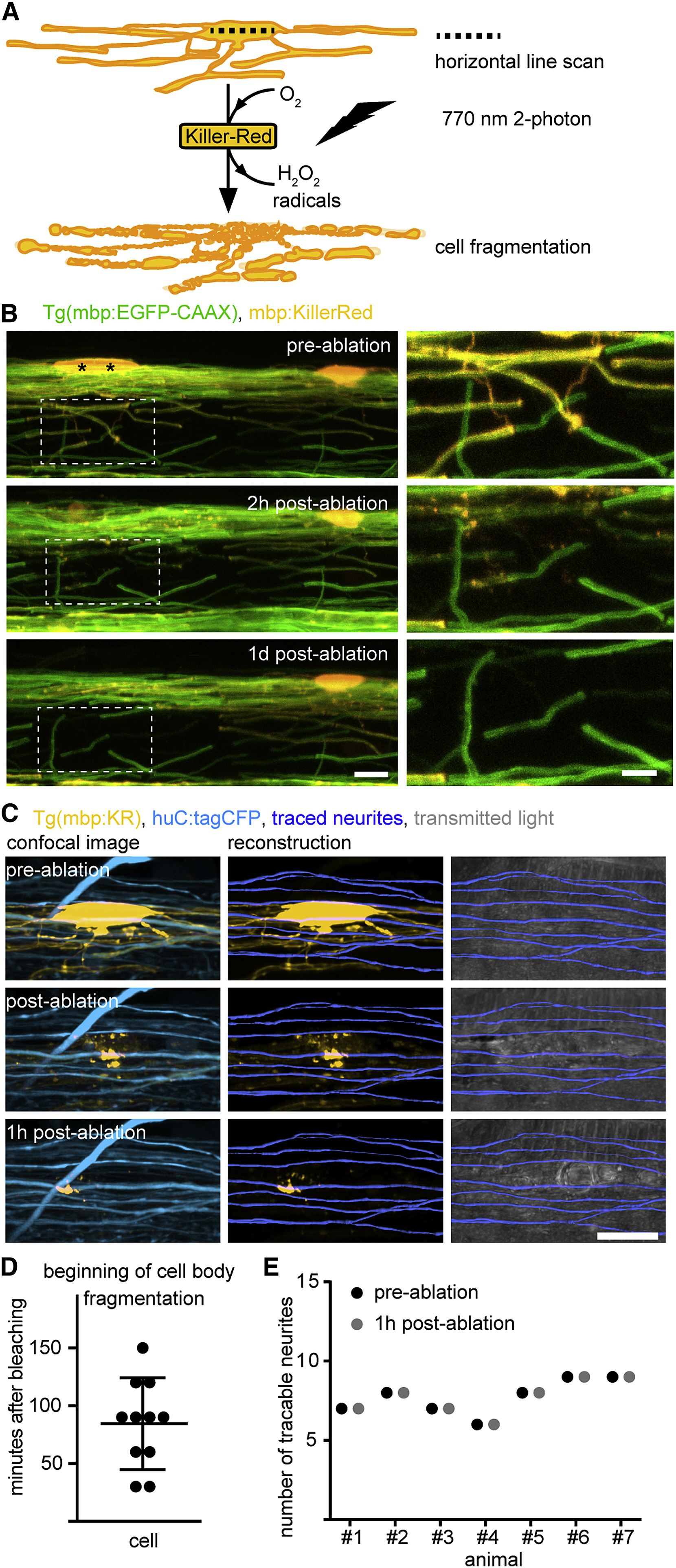
Fig. 5
Targeted Ablation of Individual Oligodendrocytes without Causing Bystander Axon Damage
(A) Schematic drawing of single oligodendrocyte ablation using two-photon bleaching of mbp:KillerRed-labeled cells.
(B) Timeline of confocal images of individual mbp:KillerRed-expressing oligodendrocytes in Tg(mbp:EGFP-CAAX) full transgenic zebrafish at 7 days post-fertilization (dpf) before and after two-photon bleaching of the cells marked by the asterisk. Scale bars, 10 μm (left), 5 μm (right).
(C) Confocal images and axon reconstructions of huC:tagCFP-labeled axons in Tg(mbp:KillerRed) zebrafish before and after oligodendrocyte ablation at 4 dpf. Scale bar, 10 μm.
(D) Quantification of the beginning of cell body disintegration after two-photon bleaching of mbp:KillerRed-expressing cells at 4 dpf. Data are expressed and mean ± SD.
(E) Quantification of traced neurites in direct proximity to the ablation site before and after oligodendrocyte ablation as shown in (C).
See also Figure S3 and Movie S3.