
|
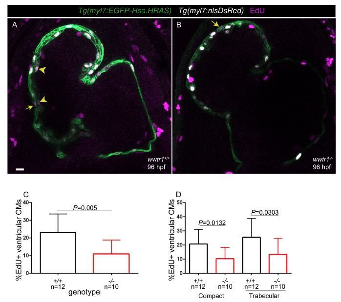
Fig. S2
Reduction in the number of EdU+ cardiomyocytes in wwtr1-/-hearts at 96 hpf.
96 hpf WT (A) and mutant (B) heats stained for EdU after 16 hours of incubation (80-96 hpf). myl7:EGFP-Hsa.HRAS and myl7:nlsDsRed expression lavel cardiomyocyte membranes and nuclei, respectively. Yellow arrows point to EdU+ compact layer cardiomyocytes. Yellow arrowheads point to EdU+ trabecular layer cardiomyocytes. Scale bar, 20 μm. (C) Quantification of EdU+ cardiomyocytes in the outer curvature of the ventricle in WT and mutant hearts. (D) Quantification of EdU+ cardiomyocytes in the compact and trabecular layers in WT and mutant hearts. Error Bars are one unit of standard deviation . P-Values were calculated by Student's t-test.