
Fig. 9
- ID
- ZDB-IMAGE-180524-21
- Publication
- Sánchez-Iranzo et al., 2018 - Tbx5a lineage tracing shows cardiomyocyte plasticity during zebrafish heart regeneration
- All Figures
- Figures for Sánchez-Iranzo et al., 2018
|
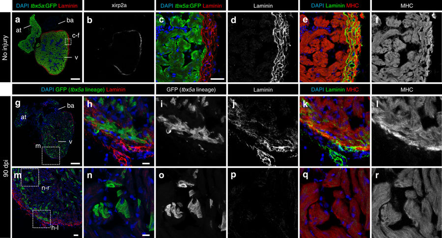
Fig. 9
Trabecular tbx5a-derived cardiomyocytes within the cortical myocardium are surrounded by Laminin. Immunofluorescence with anti-Laminin, anti-GFP, and anti-Myosin Heavy Chain (MHC) on ventricle sections. a–f Uninjured adult tbx5a:GFP ventricle. Laminin expression was observed in the cortical layer but not in the trabecular layer showing a complementary pattern with tbx5a:GFP (n = 3/3). g–r Double transgenic tbx5a:mCherry-p2a-CreERT2;ubb:loxP-lacZ-loxP-GFP adult fish were treated with 4-Hydroxytamoxifen (4-OHT) from 84 to 72 and 60 to 48 hours before cryoinjury. Hearts were fixed at 90 days postinjury (dpi). GFP+ cells marking the tbx5a lineage within the cortical layer were surrounded by Laminin staining (n = 6/6). at, atrium; ba, bulbus arteriosus; v, ventricle. Scale bars, a, g 100 µm, c, m 25 μm, and h, n 10 µm