
IMAGE
Fig. 2
- ID
- ZDB-IMAGE-180426-1
- Genes
- Publication
- Ji et al., 2017 - Involvement of Lypge in the formation of eye and pineal gland in zebrafish
- All Figures
- Figures for Ji et al., 2017
Image
|
Figure Caption
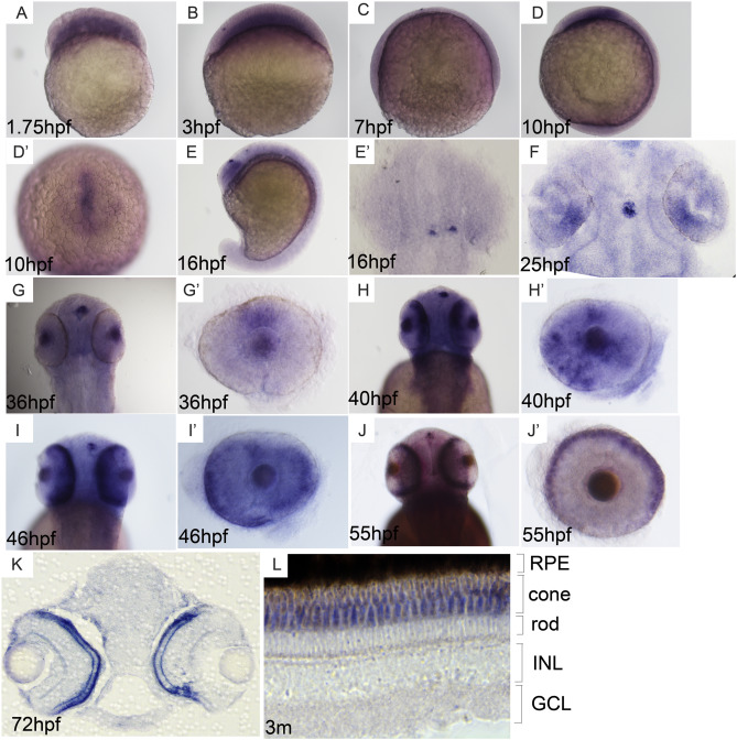
Fig. 2 Expression pattern of lypge. (A–C) Weak expression is observed from cleavage to gastrula stage. (D, D') Expression is detected in prechordal plate at bud stage. (E, E') Expression is tested in early pineal gland cells at about 16 hpf. (F–J') Expression is firstly examined in dorsal retina at about 25 hpf and in cone photoreceptor progenitor at about 40 hpf. (K–L) lypge is expressed in cone photoreceptor at 72 hpf and adult. A–D and E: the lateral view of embryos, D': animal pole view, E', F and G, H, I, J: dorsal view, G', H′, I′ and J': disconnected single eye, K and L: sectioned eye.
Figure Data
Acknowledgments
This image is the copyrighted work of the attributed author or publisher, and
ZFIN has permission only to display this image to its users.
Additional permissions should be obtained from the applicable author or publisher of the image.
Reprinted from Gene, 642, Ji, D., Wang, S., Li, M., Zhang, S., Li, H., Involvement of Lypge in the formation of eye and pineal gland in zebrafish, 491-497, Copyright (2017) with permission from Elsevier. Full text @ Gene