
|
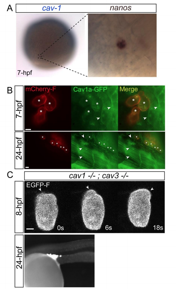
Fig. S3
The role of caveolae in blebbing. Related to Figure 4
(A) Two-color in-situ hybridization for caveolin1 (blue) and nanos3 (brown) mRNAs showing their co-expression within a PGC. (B) Global expression of Cav1a-GFP in PGCs and in somatic cells at 7hpf (upper panel) and 24hpf (lower panel), with the membrane of the PGCs labeled with mCherry-F (asterisks). Arrowheads point at membrane localization of the Cav1a-GFP to the plasma membrane of nearby somatic cells, while the PGC membrane is largely devoid of GFP signal. (C) Formation of blebs (arrowheads) at the front of a cav-1; cav-3 maternal-zygotic mutant migrating live PGC (upper panels, 8hpf) and proper arrival of the PGCs at the position where the gonad develops in 24hpf embryo of the same genotype (lower panel, 24hpf). The experiments were performed 3 times, with a total of 28 wild-type and 12 mz cav1; cav3 embryos assessed. Scale bar=5 µm.
Reprinted from Developmental Cell, 43(5), Goudarzi, M., Tarbashevich, K., Mildner, K., Begemann, I., Garcia, J., Paksa, A., Reichman-Fried, M., Mahabaleshwar, H., Blaser, H., Hartwig, J., Zeuschner, D., Galic, M., Bagnat, M., Betz, T., Raz, E., Bleb Expansion in Migrating Cells Depends on Supply of Membrane from Cell Surface Invaginations, 577-587.e5, Copyright (2017) with permission from Elsevier. Full text @ Dev. Cell