
|
Fig. S1
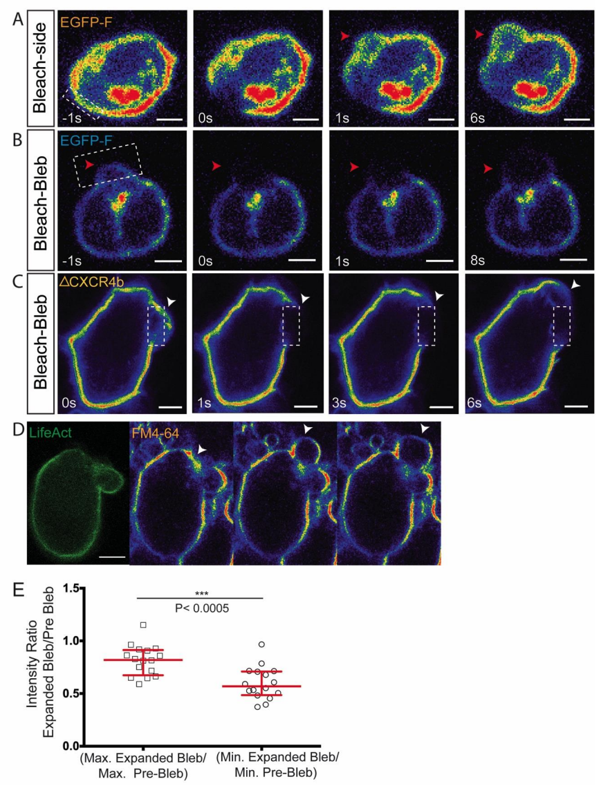
Lack of directed membrane flow during bleb formation. Related to Figure 2.
(A) An area of Farnesylated-EGFP labeled membrane adjacent to a forming bleb (arrowhead) was photobleached and the distribution of the fluorescence was assessed. Despite the expansion of the bleb, no directional material flow could be observed, 10 cells analyzed. (B) Photobleaching of Farnesylated-EGFP on the membrane of an inflating bleb (red arrowheads) reveals expansion of the dark area during bleb formation, 10 cells analyzed. (C) Photobleaching of a truncated non-internalizable, non-ligand binding form of Cxcr4b fused to EGFP. The photobleaching experiment reveals no membrane flow in the direction of the region where blebs are formed (white arrowheads), 5 cells were analyzed. (D) EGFP LifeAct–expressing PGC located within the tissue of a deyolked embryo (to allow lipid-dye labeling, see methods). The fluorescence level of the lipid dye FM4-64 observed in a single optical plane shows non-uniform signal intensity and a reduction in signal within the inflating bleb (arrowhead). (E) Normalized maximum and minimum intensity ratios of the lipid dye, FM4-64 over 3 Z-planes (maximum intensity projection) (n=16 cells). The graph shows median and interquartile range. P value was calculated using non-parametric Mann Whitney U test. Scale bars=5µm.
Reprinted from Developmental Cell, 43(5), Goudarzi, M., Tarbashevich, K., Mildner, K., Begemann, I., Garcia, J., Paksa, A., Reichman-Fried, M., Mahabaleshwar, H., Blaser, H., Hartwig, J., Zeuschner, D., Galic, M., Bagnat, M., Betz, T., Raz, E., Bleb Expansion in Migrating Cells Depends on Supply of Membrane from Cell Surface Invaginations, 577-587.e5, Copyright (2017) with permission from Elsevier. Full text @ Dev. Cell