
|
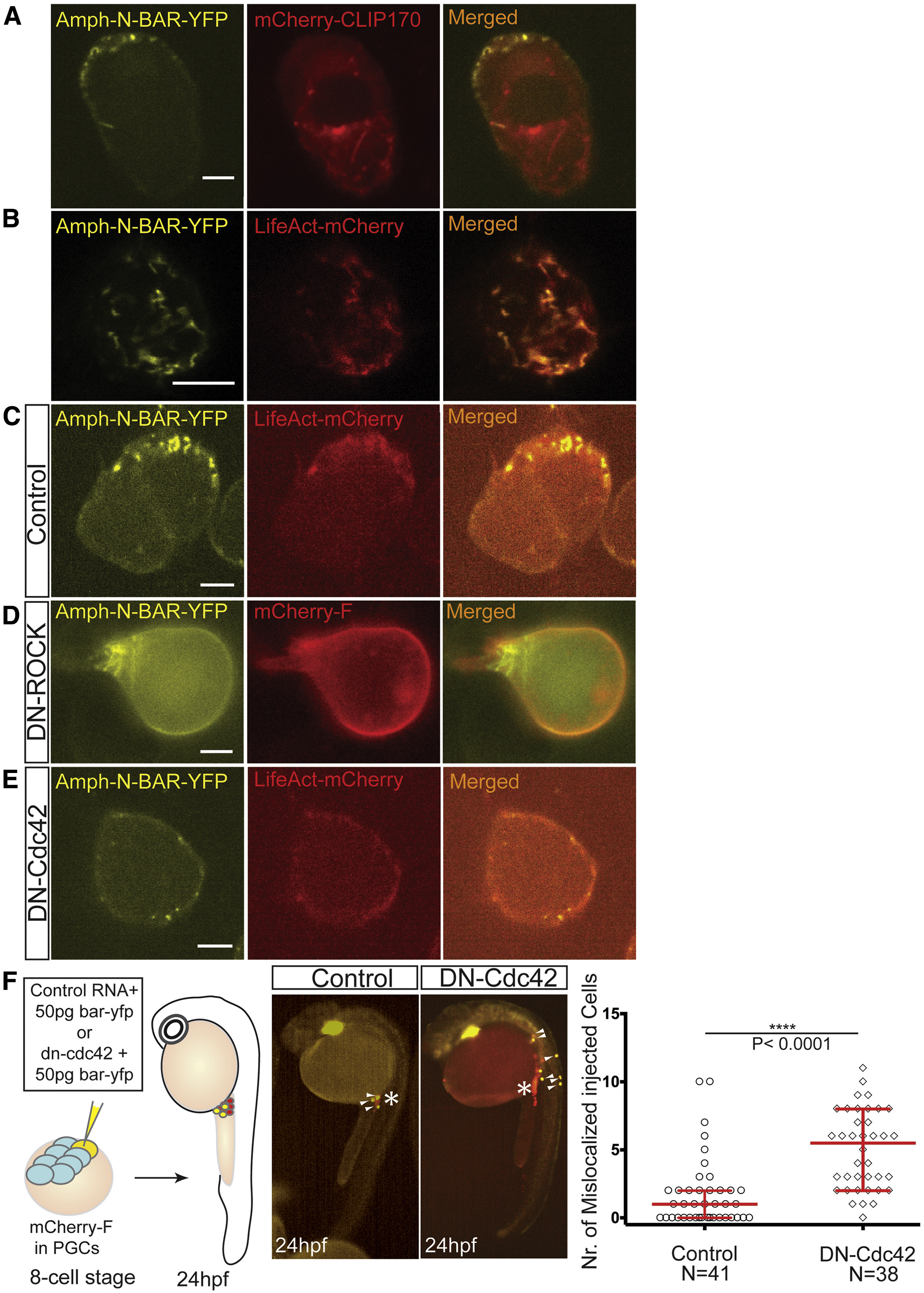
Fig. 5
Membrane invaginations in PGCs migrating within live embryos require Cdc42 for formation and colocalize with polymerized actin structures
(A) Lack of co-localization between membrane invaginations labeled by Amph-N-BAR-YFP and microtubules labeled with mCherry-CLIP170.
(B) Confocal images of membrane invaginations labeled with the Amph-N-BAR-YFP protein and their co-localization with polymerized actin labeled with LifeAct-mCherry protein. The cell orientation is such that the front is facing the lens.
(C) Co-localization of membrane invaginations in polarized PGCs labeled by Amph-N-BAR-YFP with polymerized actin labeled with LifeAct-mCherry protein. The cell front faces upper right.
(D) Membrane invaginations labeled with the Amph-N-BAR-YFP protein in cells in which contractility was inhibited by the expression of a dominant-negative form of ROCK (DN-ROCK). mCherry-F labels the plasma membrane.
(E) Dominant-negative form of Cdc42 (DN-Cdc42) expressed in the cells reduces the number of dynamic membrane invaginations labeled with Amph-N-BAR YFP. Polymerized actin is labeled with LifeAct-mCherry protein.
(F) Inhibition of Cdc42 function in a fraction of the PGC population led to mislocalization of the treated cells (yellow cells). The asterisks mark the migration target of the cells in 24 hpf embryos and the white arrowheads point to PGCs expressing a control or the DN-Cdc42 proteins. The graph shows the quantitation of the result (n = 41 and n = 38 embryos from two independent experiments). Median (1 and 5, respectively) and interquartile range are shown. The p value (∗∗∗∗p < 0.0001) was calculated using a non-parametric Mann-Whitney U test.
Scale bars, 5 μm.
Reprinted from Developmental Cell, 43(5), Goudarzi, M., Tarbashevich, K., Mildner, K., Begemann, I., Garcia, J., Paksa, A., Reichman-Fried, M., Mahabaleshwar, H., Blaser, H., Hartwig, J., Zeuschner, D., Galic, M., Bagnat, M., Betz, T., Raz, E., Bleb Expansion in Migrating Cells Depends on Supply of Membrane from Cell Surface Invaginations, 577-587.e5, Copyright (2017) with permission from Elsevier. Full text @ Dev. Cell