
|
Fig. 4
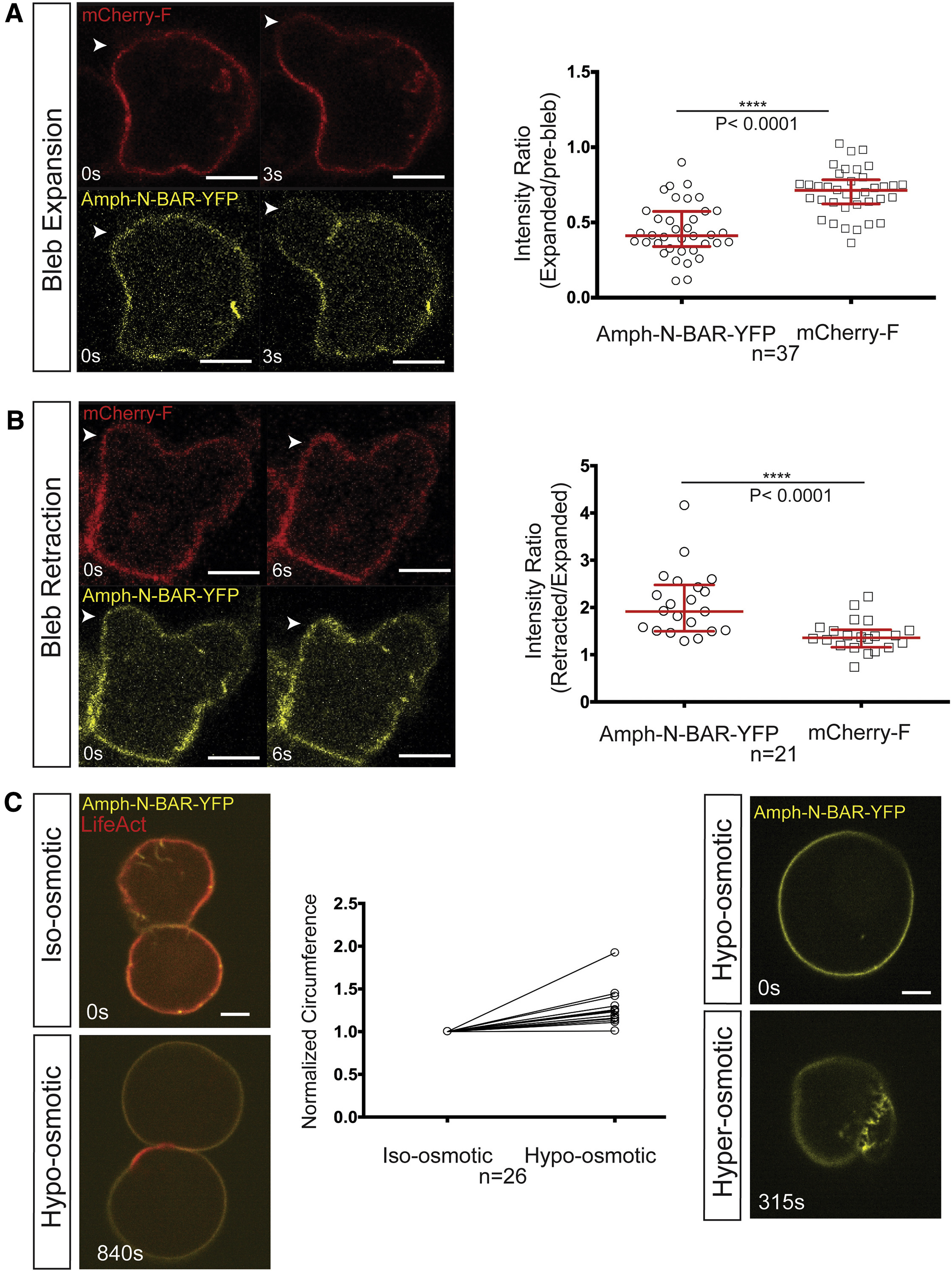
Analysis of Plasma Membrane Invaginations Unfolding and Folding during Bleb Inflation and Retraction
(A) The YFP-labeled N-BAR domain of amphiphysin interacts with the plasma membrane and marks membrane structures protruding into the cytoplasm, and the mCherry-F protein labels the PGC's plasma membrane. White arrowheads point to the position of bleb expansion. The graph on the right shows that the signal intensity of the BAR domain (Amph-N-BAR-YFP) on the plasma membrane is reduced upon bleb expansion, with a smaller reduction observed using the farnesylated mCherry (mCherry-F) probe (median = 0.41 and 0.71 for the N-BAR probe and the mCherry-F, respectively; n = 37 cells from 21 embryos).
(B) A similar analysis to that in (A) was conducted during bleb retraction. White arrowheads point to the position of bleb retraction. The graphs present the intensity ratio of each of the two probes, Amph-N-BAR-YFP and mCherry-F, between the retracted bleb and the expanded bleb (median = 1.91 and 1.35 for the N-BAR probe and the mCherry-F, respectively; n = 21 cells from 15 embryos).
(C) Two PGCs in deyolked embryos expressing N-BAR domain of amphiphysin and lifeAct in iso-osmotic (left, upper panel) and later in hypo-osmotic conditions (left, lower panel). The graph shows the change in the circumference of the cells in a confocal section after the exposure to hypo-osmotic medium. The values are normalized to the circumference in the iso-osmotic solution (n = 26 cells). Right panels show a PGC expressing N-BAR domain of amphiphysin (Amph-N-BAR-YFP) subjected to a change from hypo-osmotic to hyper-osmotic conditions (right upper and lower panels, respectively).
All graphs show median and interquartile range. The p value (∗∗∗∗p < 0.0001) was calculated using a non-parametric Mann-Whitney U test. Scale bars, 5 μm. See also Figures S3 and S4.
Reprinted from Developmental Cell, 43(5), Goudarzi, M., Tarbashevich, K., Mildner, K., Begemann, I., Garcia, J., Paksa, A., Reichman-Fried, M., Mahabaleshwar, H., Blaser, H., Hartwig, J., Zeuschner, D., Galic, M., Bagnat, M., Betz, T., Raz, E., Bleb Expansion in Migrating Cells Depends on Supply of Membrane from Cell Surface Invaginations, 577-587.e5, Copyright (2017) with permission from Elsevier. Full text @ Dev. Cell