
Fig. 7
- ID
- ZDB-IMAGE-180420-45
- Publication
- Richter et al., 2017 - Small molecule screen in embryonic zebrafish using modular variations to target segmentation
- All Figures
- Figures for Richter et al., 2017
|
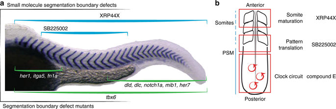
Fig. 7
Overview of selected direct segmentation phenotypes. a Schematic of axial distributions of known mutant somite boundary defects (green brackets) and newly identified small molecule perturbation defects (blue brackets, this paper). Her1, integrin α5 [itga5] and fibronectin 1a [fn1a] anterior defects; T-box 6 [tbx6] mutants, defects along axis; mutants deltaD [dld], deltaC [dlc], notch1a, mindbomb E3 ubiquitin protein ligase 1 [mib1] and her7, posterior defects11,12,71,72,73,74. b Schematic highlighting steps of segmentation (clock circuit, pattern translation, somite maturation) in red boxes and examples of small molecules from the screen that affect the corresponding step