
Fig. 2
- ID
- ZDB-IMAGE-180418-7
- Genes
- Publication
- Goody et al., 2017 - Influenza A Virus Infection Damages Zebrafish Skeletal Muscle and Exacerbates Disease in Zebrafish Modeling Duchenne Muscular Dystrophy
- All Figures
- Figures for Goody et al., 2017
|
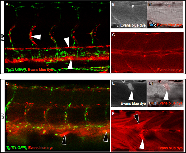
Fig. 2 IAV infection compromises the sarcolemma All embryo images are side mounts, dorsal top, anterior left. (A) Tg(fli1:GFP) zebrafish embryo with labeled endothelial cells (green) DC-injected with PBS plus EBD (red). EBD remains in the vasculature. White arrowheads point to EBD in an intersomitic vessel (top left), the dorsal aorta (middle), and the caudal vein (bottom right). (B-C) Wild-type zebrafish injected with PBS plus EBD. (B) Cropped EBD panel. (B1) Cropped EBD and brightfield panels merged. (C) EBD panel. Note that muscle fibers are impermeable to EBD in PBS-injected zebrafish. (D) Tg(fli1:GFP) zebrafish embryo DC-injected with IAV plus EBD (red). EBD leaked out of the vasculature and penetrated muscle fibers (black arrowheads). (E-F) Wild-type zebrafish injected with IAV plus EBD. (E) Cropped EBD panel. (E1) Cropped EBD and brightfield panels merged. (F) EBD panel. Note the uptake of EBD by long (black arrowheads) and retracted fibers (white arrowheads) indicative of sarcolemma damage.