
Fig. 1
- ID
- ZDB-IMAGE-180418-12
- Genes
- Publication
- Sarmah et al., 2017 - Embryonic Ethanol Exposure Affects Early- and Late-Added Cardiac Precursors and Produces Long-Lasting Heart Chamber Defects in Zebrafish
- All Figures
- Figures for Sarmah et al., 2017
|
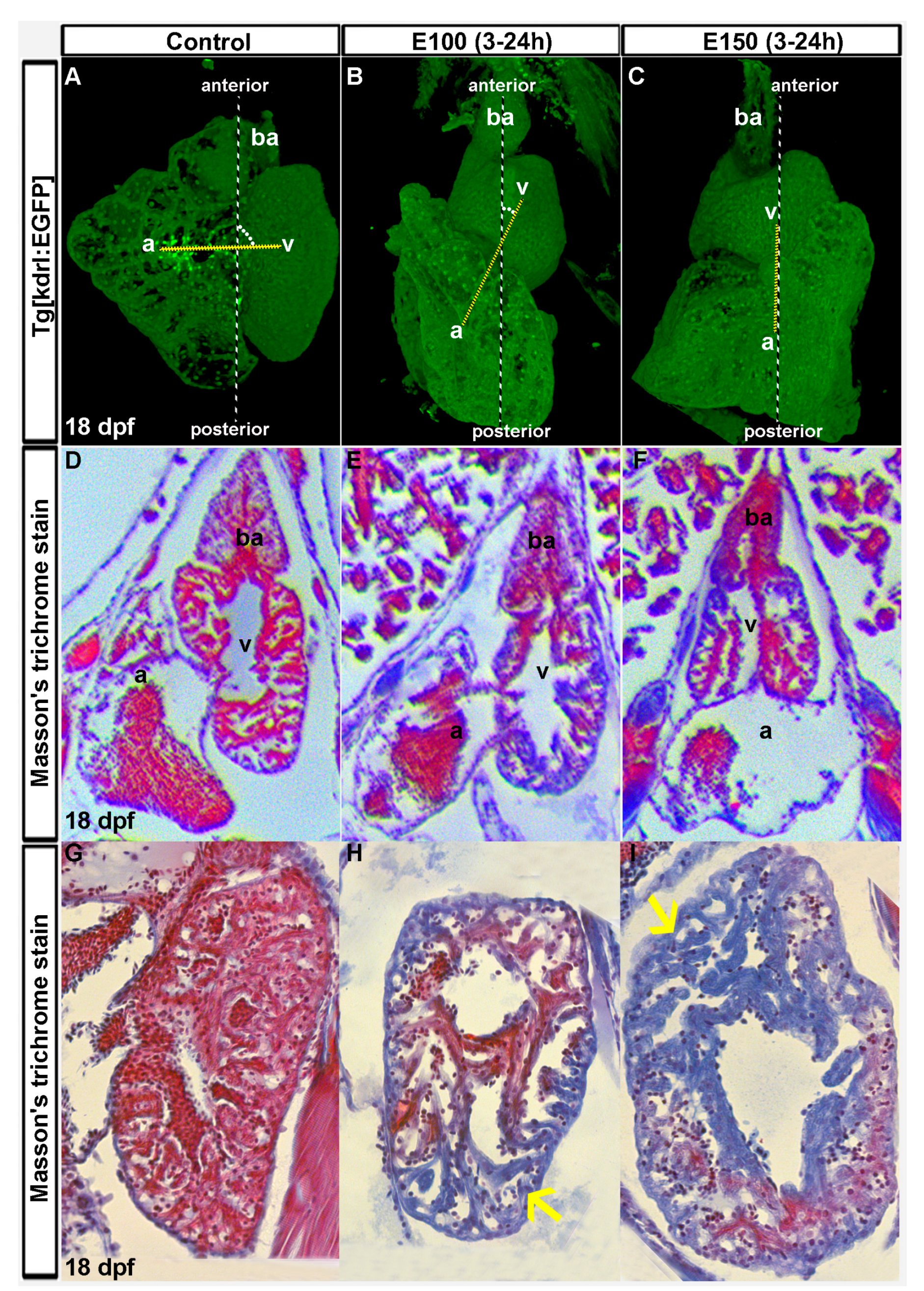
Fig. 1 Ethanol-induced cardiac chamber defects occurring during embryogenesis persisted in older zebrafish larvae. (A–C) 3D renderings of confocal sections of Tg(kdrl:EGFP) show closely attached atrium and ventricle residing side by side in the control larva (A); ventricles are on the top of the atria in ethanol-exposed larvae (B,C); Dotted yellow line represents the line through AV valves connecting atrium and ventricle. The angle between yellow line and antero-posterior axis is shown by white dots; (D–F) Masson’s trichrome stained histology sections at the atrioventricular canal region showing both atrium and ventricle revealed defective anatomy of the ethanol-exposed larvae (E,F) compared to control (D); (G–I) Masson’s trichrome stained histology sections of the ventricle showed healthy looking cardiac muscle (red colored trabeculae) in control larva (G); and myocardial damage (blue colored trabeculae, yellow arrows) in ethanol-exposed larvae (H,I).