
Image
|
Figure Caption
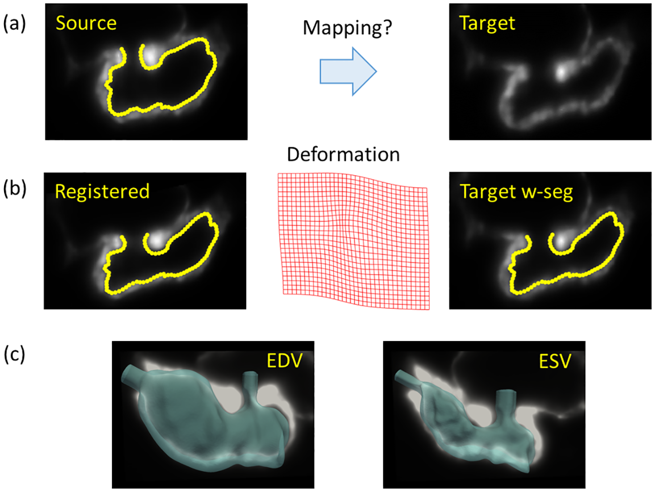
Fig. 3
Intensity-based non-rigid deformable image registration methods are used to extract ventricular endocardial motion from 4-D light sheet image data.
(a) We have chosen a source image with segmented endocardium at mid-diastole (left) and a target image at end-systole (right). (b) We perform registration using the MIRT framework to obtain the registered image (left), and compute the deformation field (middle) that is then used to morph the segmented endocardium. We note a reasonable agreement between the morphed endocardium boundary and the target image (right). (c) The registered 3D endocardial surfaces superposed on the corresponding background image are shown at end-diastolic (EDV, left) and end-systolic (ESV, right) phases of the cardiac cycle.
Acknowledgments
This image is the copyrighted work of the attributed author or publisher, and
ZFIN has permission only to display this image to its users.
Additional permissions should be obtained from the applicable author or publisher of the image.
Full text @ PLoS Comput. Biol.