
Fig. 1
- ID
- ZDB-IMAGE-180329-16
- Genes
- Publication
- Zheng et al., 2017 - Spexin Suppress Food Intake in Zebrafish: Evidence from Gene Knockout Study
- All Figures
- Figures for Zheng et al., 2017
|
Fig. 1
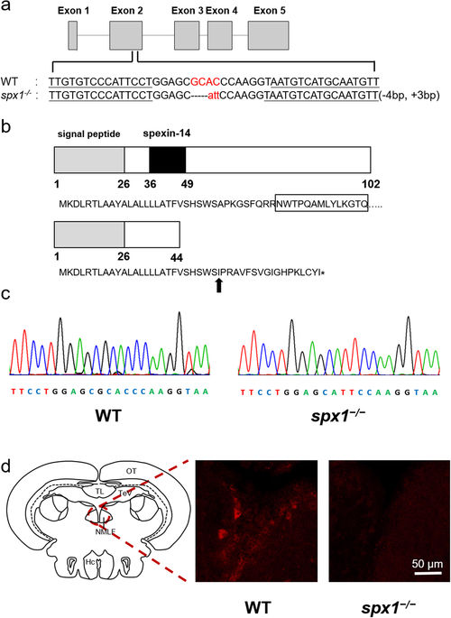
Targeted disruption of zebrafish SPX1 gene. (a) The location of the TALEN binding sites on the zebrafish SPX gene and the mutated genotype analyzed in this study. The TALEN binding sites are underlined. The inserted nucleotides are shown in lower case letters. (b) Nucleotide and amino acid sequence data for wild type and spx1 −/− genotype. Letters in box indicates the mature 14 amino acid SPX1 peptide. The new stop codon of the mutant is indicated by asterisk. Black arrow indicates the mutation starting position. (c) Comparison of two genotype sequences. (d) Detection of SPX1 expression in the brain of WT and spx1 −/− mutant fish. Note that the expression of SPX1 (red) is present in the nucleus of medial longitudinal fasciculus (NMLF) in WT while no signal can be observed in the mutant one. Hc, caudal zone of the periventricular hypothalamus, NMLF, nucleus of the medial longitudinal fascicle; OT, optic tectum; TeV, tectal ventricle; TL, torus longitudinalis.