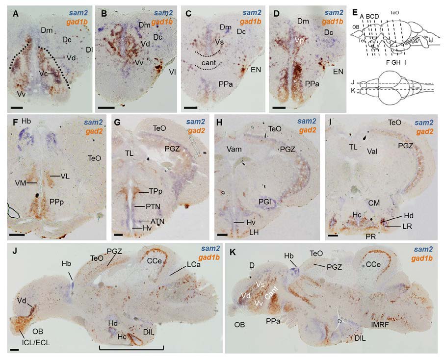
Fig. S4
Two color in situ hybridization of sam2 with gad1b or gad2 in adult brain. (A-D) Cross section of telencehpalic region. (F-I) Cross section of diencephalic area. (J, K) Sagittal section of whole brain. Expression region of sam2 is partially overlapped by that of gad1b in several brain regions, including telencephalon and hypothalamus. Levels of the sections (top, cross section; bottom, sagittal section) are indicated in (E). Scale bar, 100 μm. ATN, anterior tuberal nucleus; Cant, anterior commissure; CCe, corpus cerebelli; CM, corpus mamillare; D, area dorsalis telencephlali; Dc, central zone of area dorsalis telencephali; DIL, diffuse nucleus of the inferior lobe; Dl, lateral zone of area dorsalis telencephali; Dm, medial zone of area dorsalis telencephali; ECL, external cellular layer of olfactory bulb; EN, entopedunclular nucleus; Hc, caudal zone of periventricular hypothalamus; Hd, dorsal zone of periventricular hypothalamus; Hv, ventral zone of periventricular hypothalamus; ICL, internal cellular layer of olfactory bulb; IMRF, intermediate reticular formation; LCa, caudal lobe of the cerebellum; LH, lateral hypothalamic nucleus; LR, lateral recess of diencephalic ventricle; OB, olfactory bulb; PGl, lateral preglomerular nucleus; PGZ, periventricular gray zone; PPa, parvocelluar preoptic nucleus, anterior part; PPp, parvocellular preoptic nucleus, posterior part; PR, posterior recess; PTN, posterior tuberal nucleus; TL, torus longitudinalis; TPp, periventricular nucleus of posterior tuberculum; Val, lateral division of vlvula cerebelli; Vam, medial division of valvula cerebelli; Vc, central nucleus of area ventral telencephali; Vd, dorsal nucleus of area ventral telencephali; VL, ventrolateral thalamic nucleus; VM, ventromedial thalamic nucleus; Vp, Ventral pallium; Vs, supracommissural nucleus of area ventral telencephali; Vv, ventral nucleus of area ventral telencephali.

