
Fig. S3
- ID
- ZDB-IMAGE-180207-11
- Publication
- Choi et al., 2018 - Targeted knockout of a chemokine-like gene increases anxiety and fear responses
- All Figures
- Figures for Choi et al., 2018
|
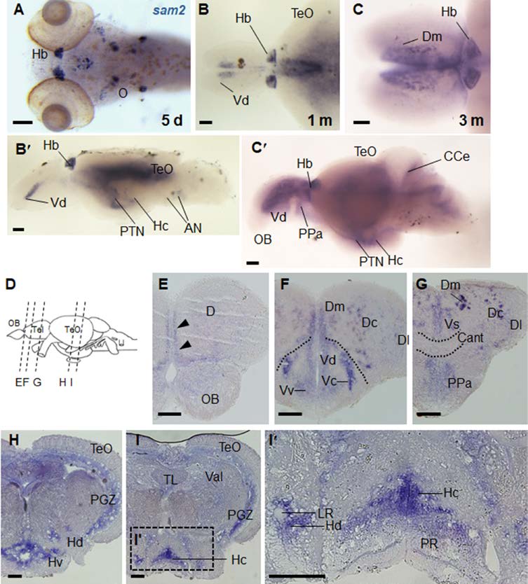
Fig. S3
Expression of sam2 in the larval and adult brain. Whole-mount in situ hybridization of sam2 in larva and adult (1- and 3-month; 1 m, 3 m) (A) sam2-expressing cells in 5 day-old larvae. Habenula (Hb) expression is already prominent at this stage. Expression in the otic neurons (O) is also seen. (B, B’) At 1-month, expression of sam2 in the brain. Dorsal telencephalic view (B) and lateral view (B’). (C, C’) sam2 expression in the adult brain. Dorsal telencephalic view (C) and lateral view (C’). (E-I’) Serial sections of adult brain. Levels of the cross-sections are indicated in (D). Scale bar, 100 μm. AN, auditory nerve; Cant, anterior commissure; CCe, corpus cerebelli; D, area dorsalis telencephlali; Dc, central zone of area dorsalis telencephali; Dl, lateral zone of area dorsalis telencephali; Dm, medial zone of area dorsalis telencephali; Hc, caudal zone of periventricular hypothalamus; Hd, dorsal zone of periventricular hypothalamus; Hv, ventral zone of periventricular hypothalamus; LR, lateral recess of diencephalic ventricle; OB, olfactory bulb; PGZ, periventricular gray zone; PPa, parvocelluar preoptic nucleus, anterior part; PR, posterior recess; PTN, posterior tuberal nucleus; TeO, optic tectum; TL, torus longitudinalis; Val, lateral division of vlvula cerebelli; Vc, central nucleus of area ventral telencephali; Vd, dorsal nucleus of area ventral telencephali; Vs, supracommissural nucleus of area ventral telencephali; Vv, ventral nucleus of area ventral telencephali.