
Image
|
Figure Caption
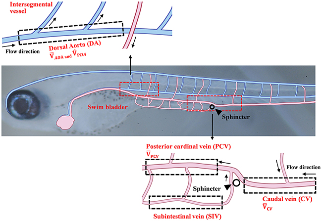
Fig. 1
Schematics of the vascular network of a 10 dpf zebrafish. Vessels are composed of the dorsal artery (DA), posterior cardinal vein (PCV), caudal vein (CV), and subintestinal vein (SIV). Each region of interest (ROI) is represented by dotted box. The time-averaged RBC velocity and peak systolic RBC velocity in the DA are defined as and , respectively. The time-averaged RBC velocities in the PCV and CV regions are defined as
Acknowledgments
This image is the copyrighted work of the attributed author or publisher, and
ZFIN has permission only to display this image to its users.
Additional permissions should be obtained from the applicable author or publisher of the image.
Full text @ Oncotarget