
Image
|
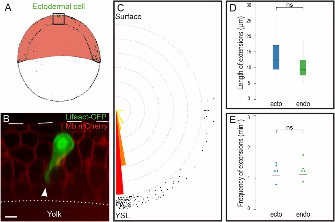
Figure Caption
Fig. S4
Ectodermal cells are protrusive and polarized. (A) Position of cell transplantation just beneath the EVL, at the animal pole of the embryo. (B–E) Ectodermal cells expressing Lifeact-GFP emit large cytoplasmic extensions (arrowhead in B) oriented toward the YSL (C), of the same length (D) and at the same frequency (E) as endodermal cells. nendo = 72 extensions, necto = 71 extensions, Pangle endo/ecto = 0.97. (Scale bar: 10 µm.) ns, nonsignificant (P > 0.05).
Acknowledgments
This image is the copyrighted work of the attributed author or publisher, and
ZFIN has permission only to display this image to its users.
Additional permissions should be obtained from the applicable author or publisher of the image.
Full text @ Proc. Natl. Acad. Sci. USA