
Image
|
Figure Caption
Fig. 4
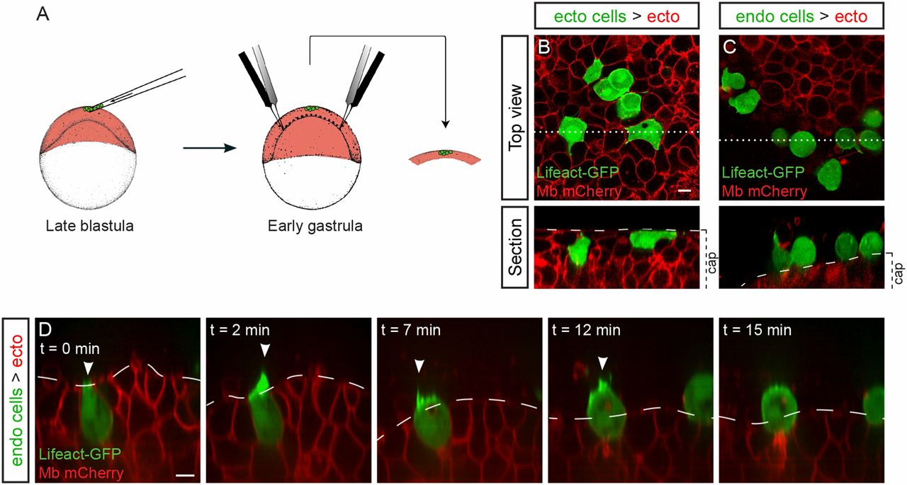
Endodermal cells are not attracted to the YSL. (A) Ectodermal or endodermal cells were transplanted to the animal pole of a host embryo. An animal cap explant was then dissected and cultured. (B and C) After 3 h of culture, transplanted ectodermal cells are still in the cap explant (B), while endodermal cells have exited (C). (B and C, Upper) Top view. (B and C, Lower) Section along the dotted line in the upper view; brackets show the position of the animal cap. (D) Endodermal cells migrate out of the explant (Movie S11) using actin-rich protrusions pointing outwards (arrowheads). (Scale bars: 10 µm.)
Acknowledgments
This image is the copyrighted work of the attributed author or publisher, and
ZFIN has permission only to display this image to its users.
Additional permissions should be obtained from the applicable author or publisher of the image.
Full text @ Proc. Natl. Acad. Sci. USA