
Fig. 8
- ID
- ZDB-IMAGE-180105-8
- Publication
- Yang et al., 2017 - Serotonin Activated Hepatic Stellate Cells Contribute to Sex Disparity in Hepatocellular Carcinoma
- All Figures
- Figures for Yang et al., 2017
|
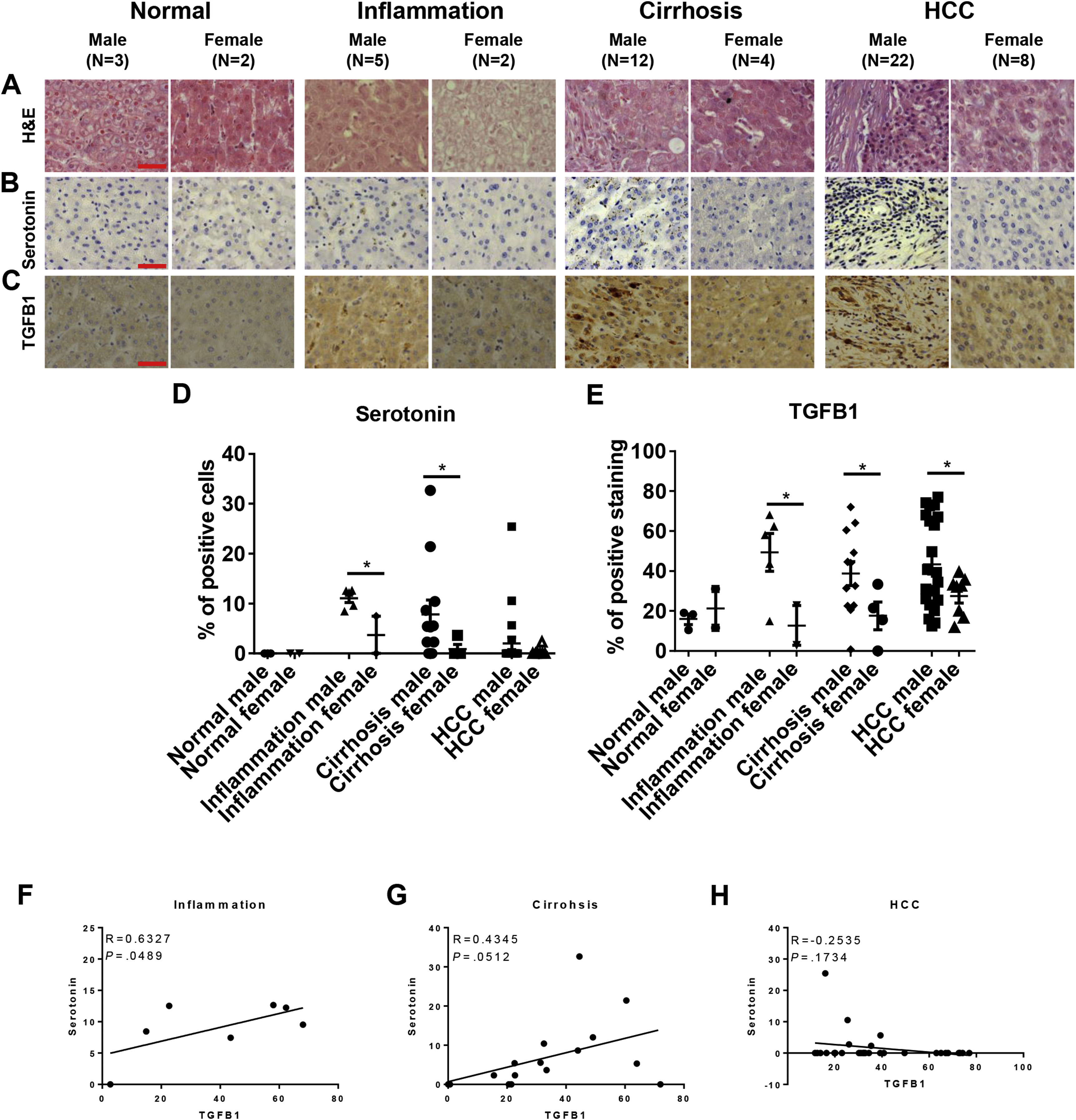
Fig. 8
Sex difference in serotonin and TGFB1 levels in human liver disease samples. A panel of liver disease samples from human patients was examined for histology by H&E staining and for serotonin and TGFB1 levels by antibody staining. These samples were categorized into normal, inflammation, cirrhosis, and HCC for both males and females. (A) H&E staining of human liver disease samples. (B and C) IHC staining of antibody against (B) serotonin and (C) TGFB1. (D and E) Quantification of the percentages of (D) serotonin or (E) TGFB1-positive liver cells in inflammation, cirrhosis, and HCC patients. (F–H) Quantification of correlation between serotonin with TGFB1 in (F) inflammation, (G) cirrhosis, and (H) HCC patients. *P < .05. Scale bar: 20 μm.