
Fig. 4
- ID
- ZDB-IMAGE-180104-4
- Genes
- Publication
- Djenoune et al., 2017 - The dual developmental origin of spinal cerebrospinal fluid-contacting neurons gives rise to distinct functional subtypes
- All Figures
- Figures for Djenoune et al., 2017
|
Fig. 4
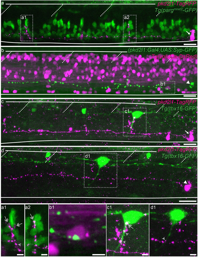
Ventral and dorsal CSF-cNs project onto distinct neuronal populations. (a–d) Z projection stacks showing contact from ventral and dorsal CSF-cNs onto different spinal targets. (a) Lateral view of a ventral CSF-cN (magenta, arrow) contacting 2 CaP motor neurons (identified based on their location within the segment) labelled in green in the Tg(parg mnet2 -GFP) transgenic line (a1,a2, double headed arrows). (b) Dorsal CSF-cN (green, arrowhead) contacting a putative V0-v interneuron (magenta, based on its dorso-ventral and lateral location) in the Tg(vglut2a:DsRed) transgenic line. (c,d) Ventral (c, arrow) and dorsal (d, arrowhead) CSF-cNs (magenta) contact CoPA sensory interneurons (green) labelled in the Tg(tbx16-GFP) transgenic line. Boxes with close-ups highlight contacts between the CSF-cN and its target. Scale bar = 20 µm (a–d) and 5 µm (a1–d1).