
|
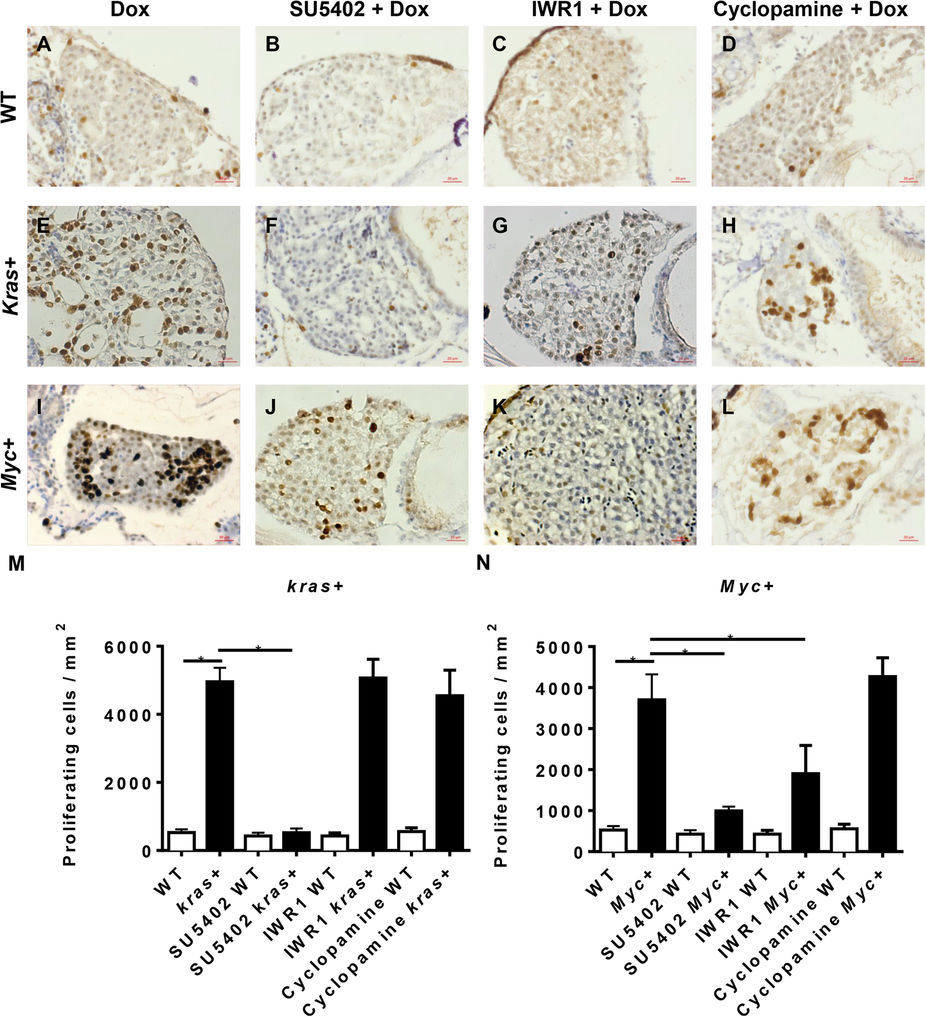
Fig. 4
Cell proliferation analysis of krasV12- and Myc-induced carcinogenesis.
7 dpf wild type (WT), kras+ or Myc+ larvae were treated with 10 μM SU5402, 10 μM IWR1 or 10 μM cyclopamine in the presence of 10 μg/ml Dox. Cell proliferation was analyzed by immunohistochemical staining with PCNA primary antibody. (A–D) Representative liver image of 7 dpf WT larvae. (E–H) Representative liver image of 7 dpf kras+ larvae. (I–L) Representative liver image of 7 dpf Myc+ larvae. (M) Statistical analysis of numbers of proliferating cells in the livers of kras+ larvae. (N) Statistical analysis of numbers of proliferating cells in the livers of Myc+ larvae. N = 20 from each groups; statistical significance: *p < 0.05, Scale bar = 20 μm.