
|
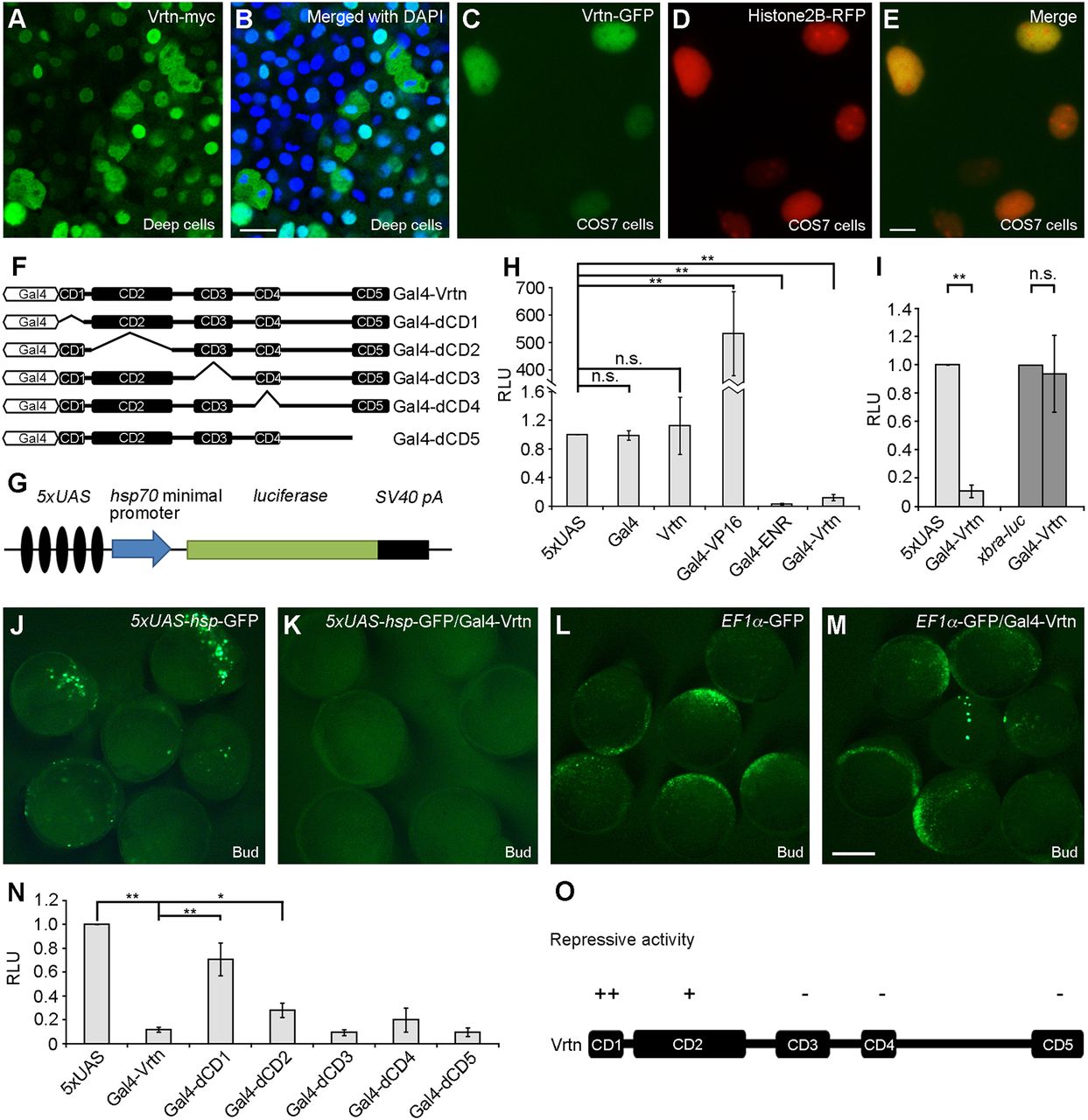
Fig. 7
Vrtn functions as a transcriptional repressor. (A,B) Nuclear localisation of Vrtn-myc in deep cells of zebrafish embryos at 50% epiboly. (C-E) Nuclear localisation of Vrtn-GFP in COS7 cells. (F) Schematic of full-length and truncated Vrtn fused with a Gal4 DNA-binding domain. (G) Schematic shows the 5×UAS-hsp-luc reporter. (H) Graph shows the specificity and validity of the one-hybrid assay system. Data are mean±s.d. from three independent experiments. (I) Gal4-Vrtn has no effect on the xbra promoter. Data are mean±s.d. from three independent experiments. (J-M) Gal4-Vrtn suppresses GFP transcription driven by the 5×UAS-hsp promoter, but not by the EF1α promoter. The embryos were injected with 5×UAS-hsp-GFP plasmid (100 pg) or EF1α-GFP plasmid (20 pg), either alone or together with Gal4-Vrtn mRNA (200 pg). (N) Graph shows the transcriptional repressor activity of full-length and truncated Vrtn. Data are mean±s.d. from three independent experiments. (O) Summary of the repressive activity of Vrtn domains. Scale bars: 20 μm in A,B; 10 µm in C-E; 200 µm in J-M.