
Fig. S2
- ID
- ZDB-IMAGE-171207-35
- Publication
- Lu et al., 2015 - Direct regulation of p53 by miR-142a-3p mediates the survival of hematopoietic stem and progenitor cells in zebrafish
- All Figures
- Figures for Lu et al., 2015
|
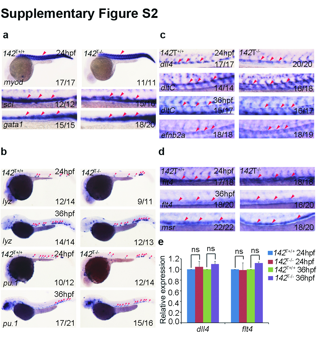
Fig. S2
Normal primitive hematopoiesis and vascular development in miR-142a knock out mutants.
(a) 142T-/- embryos displayed normal expression of primitive hematopoiesis marker scl and erythrocyte marker gata1 by WISH at 24 hpf. Somitic marker myod served as control. (b) 142T-/- embryos showed normal expression of primitive myeloid marker lyz and pu.1 by WISH at 24 and 36 hpf respectively. (c) Arterial marker dll4 dltC and efnb2a were unaffected in 142T-/- embryos by WISH at 24 and 36 hpf respectively. (d) Venous marker flt4 and msr were unaffected in 142T-/- embryos by WISH at 24 and 36 hpf respectively. (e) 142T-/- embryos showed normal expression of dll4 and flt4 at 24 and 36 hpf by qPCR (mean±SD, n=3, ns stands for no significance).