
Fig. 8
- ID
- ZDB-IMAGE-171201-34
- Genes
- Antibodies
- Publication
- Khatri et al., 2016 - Down-regulation of coasy, the gene associated with NBIA-VI, reduces Bmp signaling, perturbs dorso-ventral patterning and alters neuronal development in zebrafish
- All Figures
- Figures for Khatri et al., 2016
|
Fig. 8
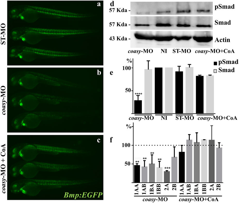
Analysis of Bmp activity in coasy-morphants.
Representative images of Tg(Bmp:EGFP) embryos injected with ST-MO (a) or coasy-MO (1.2 pmol/embryo, b and c), eventually exposed to CoA in fish water (c), at 60 hpf. A net reduction of the fluorescence intensity is evident in coasy-morphants. Results are from one representative experiment with at least 25 embryos out of two independent replicates. (d) Western blotting analysis of the amount of phosporylated Smad-5, total Smad, and Actin in embryos injected with coasy-MO (±CoA) or ST-MO (1.2 pmol/embryo), and not-injected (NI). Full size images of the gels are shown in Supplementary Fig. S7) The graph shows the quantitative analysis of the immunoblotting. (f ) Real-time RT-PCR quantification of different Bmp receptor mRNAs in morphants at 48 hpf, eventually treated with CoA in fish water. Mean values are form three independent experiments and expressed as percentage of the value in ST-MO injected embryos.