
Image
|
Figure Caption
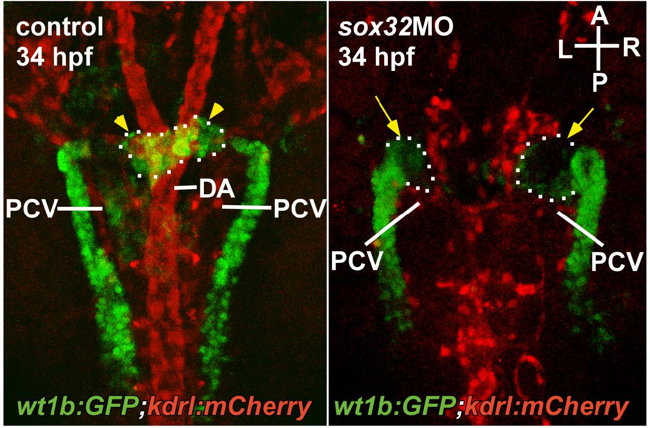
Fig. S2
Angiogenesis of the PG was defective in the sox32 morphant. The pronephric kidney and the blood vessels are delineated by green and red fluorescence respectively in the Tg(wt1b:GFP);Tg(kdrl:mCherry)ci5 embryo at the stage of 34 hpf. The relative spatial distribution of the PG (yellow arrowheads), the DA and the PCV is shown in a dorsal view. Yellow arrows indicate defective angiogenesis of the PG. A, anterior; P, posterior; R, right; L, left.
Acknowledgments
This image is the copyrighted work of the attributed author or publisher, and
ZFIN has permission only to display this image to its users.
Additional permissions should be obtained from the applicable author or publisher of the image.
Full text @ Sci. Rep.