
|
Fig. 5
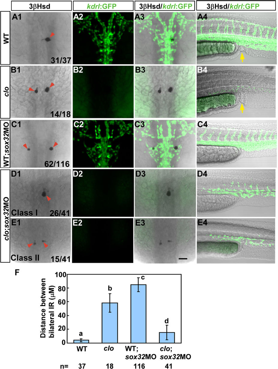
The defective interrenal migration in the sox32 morphant is suppressed by the clom39 mutation.
The sox32MO was injected into the Tg(kdrl:EGFP)s843; clom39 mutant (D1–D4,E1–E4) and its wild-type sibling (C1–C4) to inhibit endoderm formation, as verified by the absence of anal openings (C4–E4). By contrast, the clom39 mutant (B1–B4) and its wild-type sibling (A1–A4) showed normal formation of anal openings (yellow arrows in A4 and B4). All images are single confocal sections showing the IR as detected by 3β-Hsd staining (A1–E1,A3–E3) and the vascular pattern at the midtrunk (A2–E2,A3–E3) and the lower trunk (A4–E4) revealed by GFP in Tg(kdrl:EGFP)s843 at 34 hpf. In clom39, the endothelial fluorescence was absent at the midtrunk and disrupted at the lower trunk. The severe disruption of interrenal migration in the sox32 morphant was rescued when the endothelium was simultaneously deleted by the clom39 mutation, as revealed in both class I (D1–D3) and class II (E1–E3) double loss-of-function embryos. The effects of various treatments on the convergence of bilateral IRs are quantified in (F). Histograms with different letters above them are significantly different (ANOVA and Duncan’s multiple test, P < 0.05). Scale bar, 50 μm.