
|
Fig. 2
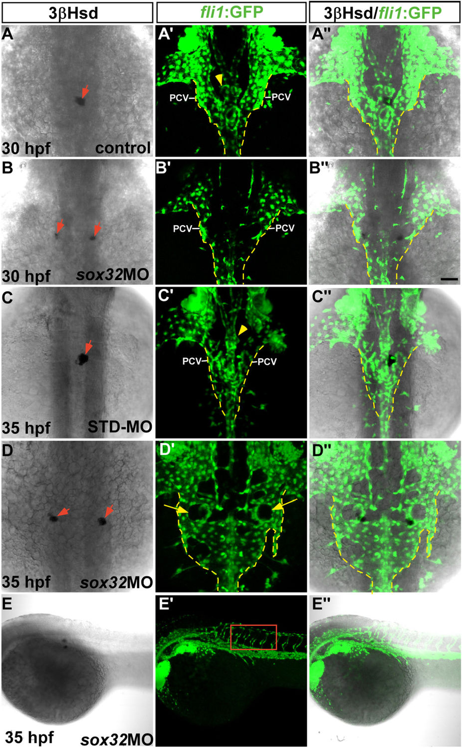
The morphology of the axial vasculature in the vicinity of the kidney and the IR in the sox32 morphant.
3β-Hsd activity staining was performed on the sox32MO-injected Tg(fli1:EGFP)y1 embryos (B–B'',D–D'',E–E'') and the uninjected (A–A'') or STD-MO-injected (C–C'') controls. All panels except (E–E'') are dorsal views with anterior oriented toward the top, while (E–E'') are dorsolateral views with anterior toward the left. The outlined area in E’ highlights the normal growth of the intersegmental vessels in the sox32 morphant. Yellow broken lines demarcate lateral boundaries of the PCV. Red arrows indicate IRs. Yellow arrowheads indicate PGs. Yellow arrows denote the pronephric tissues deficient in angiogenesis. Scale bar, 50 μm.