
|
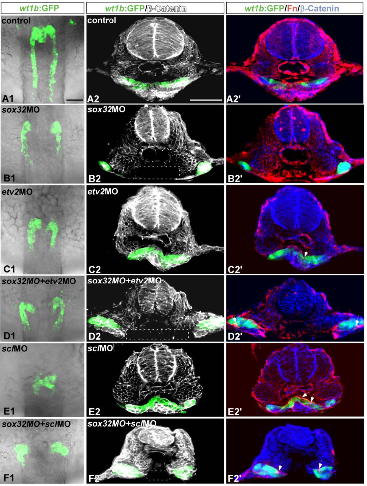
Fig. S9
The defective midline convergence of the kidney in the sox32 morphant prior to interrenal differentiation was not suppressed by either etv2MO or sclMO. sox32MO, etv2MO, sclMO, sox32/etv2 double-MOs, sox32/scl double- MOs and STD-MO were injected into Tg(wt1b:GFP) embryos to test their effects on kidney morphology at 24 hpf. (A1-F1) The kidney morphology was delineated by the expression of wt1b:GFP in the dorsal whole-mount views of the midtrunk. (A2-F2) Cross-sections showing the expression of wt1b:GFP and β-Catenin (white). (A2'-F2') Cross-sections showing the expression of wt1b:GFP, Fn (red) and β-Catenin (blue). The loss of β-Catenin in the ventral region is highlighted by white rectangles, and Fn deposits are marked by white arrows. The images shown are representative of 3, 15, 20, 16, 20 and 11 samples for control embryos, sox32 morphants, etv2 morphants, etv2/sox32 double morphants, scl morphants and scl/sox32 double morphants, respectively. Scale bar, 50 μm.