
|
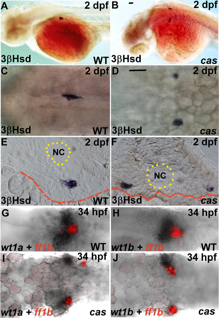
Fig. 1
The phenotypes of the PG and the IR in the cas mutant and its wild-type sibling.
(A–F) The steroidogenic IR detected by whole-mount 3β-Hsd staining at 2 dpf in the cas mutant (B,D,F) and its wild-type sibling (A,C,E). Dorsolateral (A,B) and dorsal (C,D) views are oriented with the anterior toward the left. (E,F) Cryosections with dorsal oriented toward the top. (G–J) Expression of ff1b and wt1a (G,I) or wt1b (H,J) were detected simultaneously in the cas mutant (I,J) and its wild-type sibling (G,H). The boundaries of the yolk and the notochord (NC) are highlighted by red dashed and dotted lines, respectively. Scale bar, 50 μm.