
Image
|
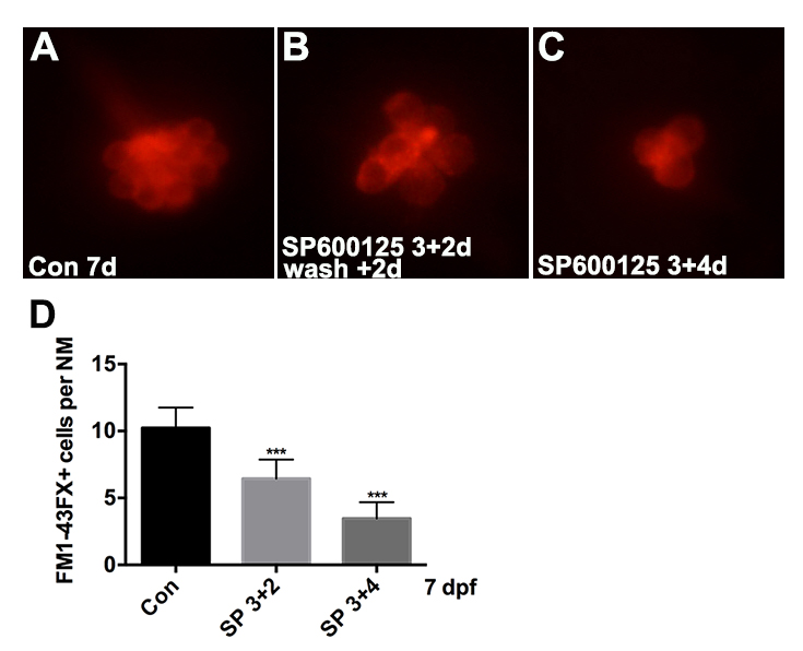
Figure Caption
Fig. S4
Effects of varying duration of SP600125 exposure on hair cell number during the period of embryonic development. (A) Control group; (B) larvae at 3 dpf were treated with 10μM SP600125 for 4 days; (C) larvae at 3 dpf were treated with 10μM SP600125 for 2 days, after which the inhibitor was washed out and hair cells were analyzed after another 2 days. (D) Quantification of FM1-43FX+ hair cells in the neuromast (NM) for each experimental condition [One-way ANOVA; F(2, 117) = 234.9, p < 0.001]. Bars are mean ± SD. n = 36-44 neuromasts per treatment. ***p < 0.001.
Acknowledgments
This image is the copyrighted work of the attributed author or publisher, and
ZFIN has permission only to display this image to its users.
Additional permissions should be obtained from the applicable author or publisher of the image.
Full text @ Front. Cell. Neurosci.