
|
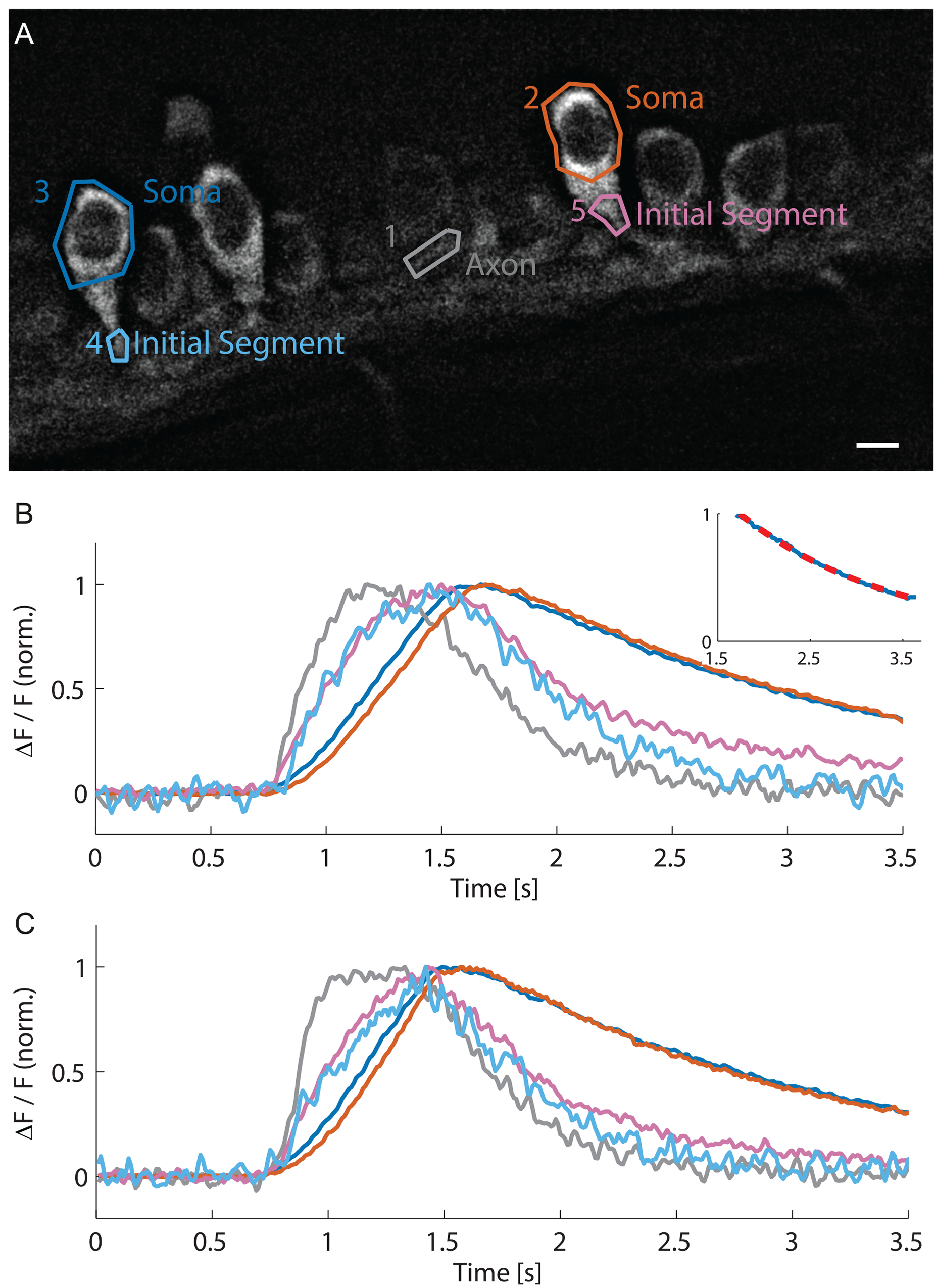
Fig. 5
Fast calcium imaging at 100 fps with HiLo microscopy reveals different dynamics in soma and initial segment.
(A) HiLo image of motor neurons expressing GCaMP5G with ROIs. Scale bar 5 μm. (B) Time course of calcium signals in soma, initial segment and axon, recorded at 100 fps. In the axon (ROI 1, gray) and the initial segments (ROI 4, light blue; ROI 5, magenta) a much faster rise is observed than in the somata (ROI 2, orange; ROI 3, dark blue). Somatic signals show a linear rise and a mono-exponential decay (Inset: blue somatic signal with red dotted fit, decay time 1.7 s, R2 = 0.998). (C) The kinetics of calcium transients in different compartments were stable over time as shown with a recording performed 2.5 min later.