
Image
|
Figure Caption
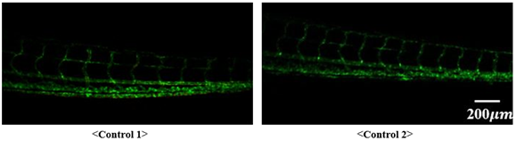
Fig. S2
Confocal microscope images of two control zebrafish models.
Control experiments using zebrafish fed normal diet for 10 days were conducted (the same amount of fluorescent cholesteryl ester without additional cholesterol). As shown in the figure below, deposition of lipids on blood vessels was not observed in the confocal microscope images of the control group.
Acknowledgments
This image is the copyrighted work of the attributed author or publisher, and
ZFIN has permission only to display this image to its users.
Additional permissions should be obtained from the applicable author or publisher of the image.
Full text @ PLoS One