
|
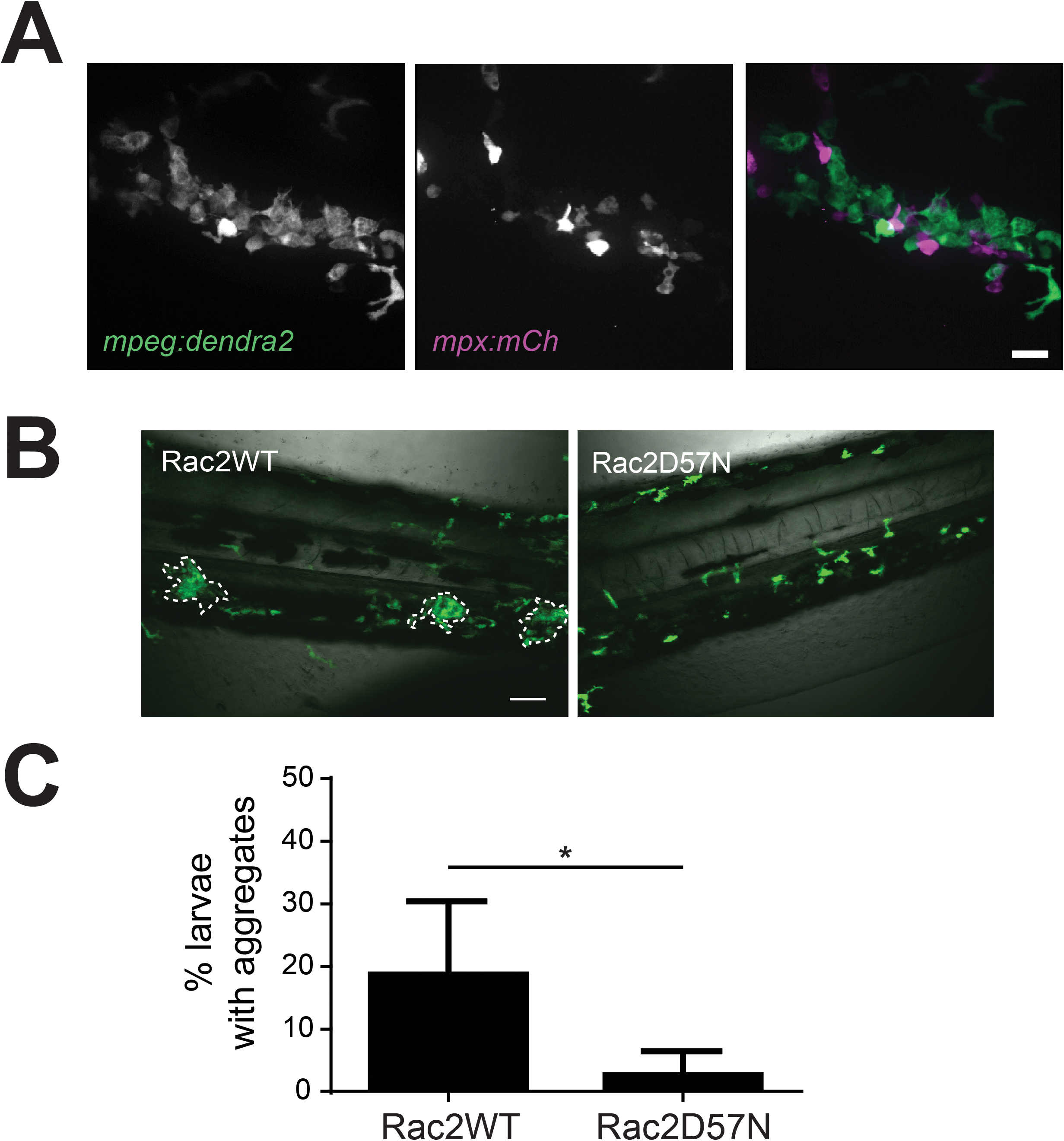
Fig. 4
Neutrophil crosstalk drives macrophage aggregate formation.
(A) Double transgenic Tg(mpeg:dendra2) x Tg(mpx:mCherry) larvae show that neutrophils are present in and around macrophage aggregates. Images at 40X; scale bar is 20 μm. (B) Representative 20X images of Rac2WT or Rac2D57N larvae at 24 hpi following inoculation. Tg(mpx:mCherry-2a-rac2wt) (Rac2WT) or Tg(mpx:mCherry-2a-rac2d57n) (Rac2D57N) were crossed to Tg(mpeg1:dendra2) and the resulting double transgenic larvae were infected with 50 CFU WT S. iniae or mock-infected with PBS. Rac2D57N larvae are defective for aggregate formation. Scale bar is 80 μm. (C) Quantification of the average total percent of larvae forming macrophage aggregates from (B).