
|
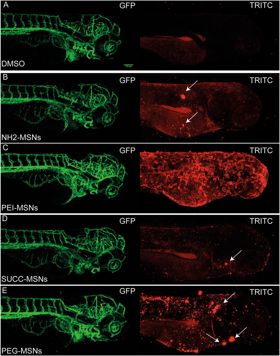
Fig. 5
Surface functionalization affects aggregation of MSNs on the embryo skin. The uptake of the nanoparticles from the aquaeous medium into the embryo was analyzed by mounting anesthesized living embryos into agarose and imaged using confocal microscopy (Leica SP5 Matrix). A transgenic strain (kdrl:EGFP) expressing a GFP labeled vasculature were used 24hpf embryos were incubated 48 h with the highest tolerated dose of the different nanoparticles e.g 100 μg/ml NH2-, SUCC- and PEG-MSNs, and 10 ug/ml of PEI-MSNs. DMSO treated embryos were used as controls and show background fluoresence (A). Maximum Z-projections are shown. The white arrows mark nanoparticle clusters on the superficial layer of the embryo.