
Fig. 4
- ID
- ZDB-IMAGE-170922-42
- Publication
- Xue et al., 2017 - The Vascular Niche Regulates Hematopoietic Stem and Progenitor Cell Lodgment and Expansion via klf6a-ccl25b
- All Figures
- Figures for Xue et al., 2017
|
Fig. 4
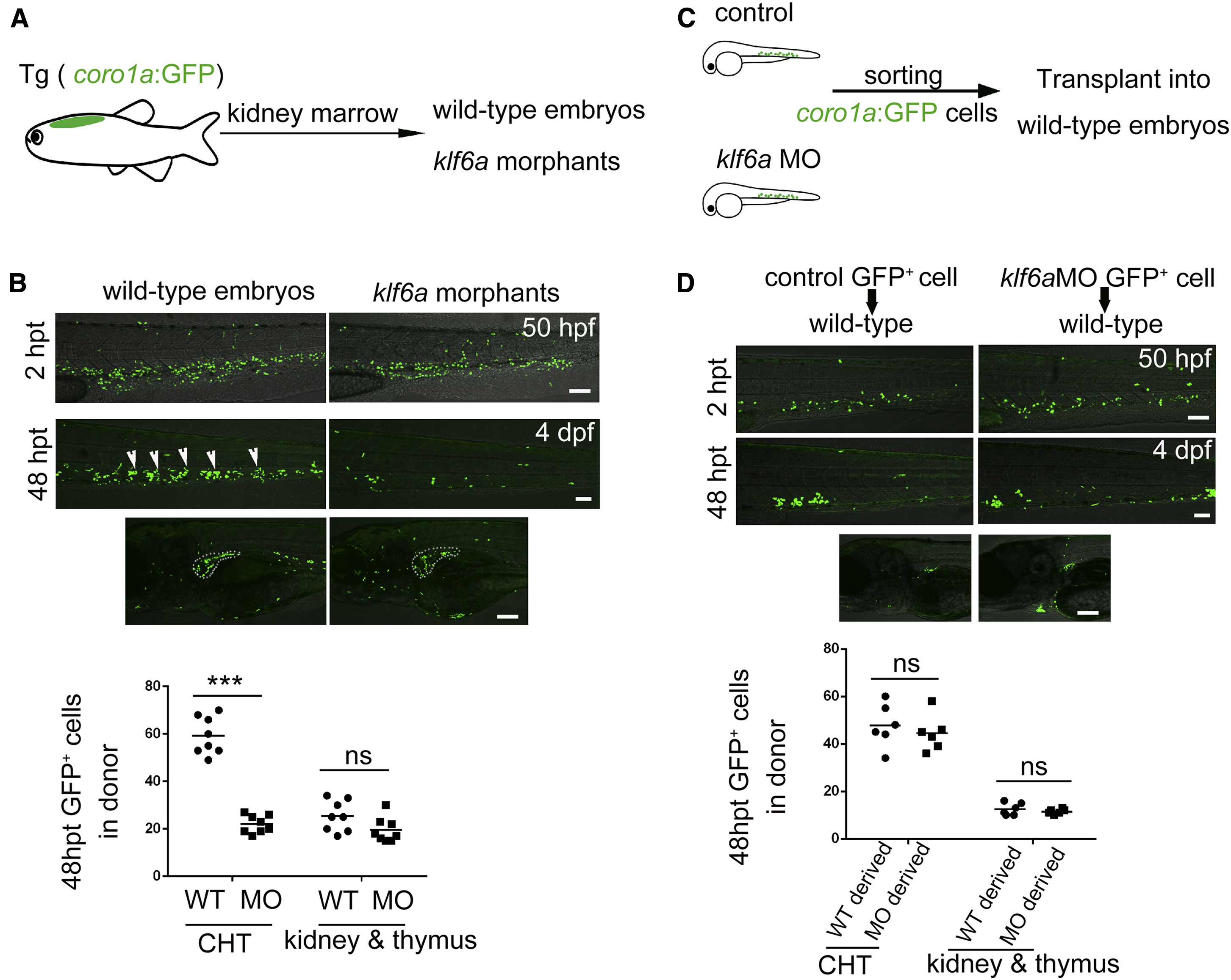
Klf6a Regulates HSPCs in the CHT Non-Cell Autonomously
(A) The model of experimental procedure of kidney marrow transplantation.
(B) Confocal imaging showing donor-derived coro1a+ cell in recipients 2 hr and 48 hr post transplantation (hpt). The statistical data show that klf6a morphants had fewer donor cells in the CHT, but there was no difference in the thymus and kidney in control and klf6a morphant recipients (mean ± SD, t test; ∗∗∗p < 0.001, ns [not significant] > 0.05, n = 8). White arrowheads indicate the coro1a+ cells. Scale bars, 50 μm.
(C) The embryos-to-embryos transplantation model.
(D) Transplantation results shows that there was no difference in the number of donor cells from either control or klf6a morphants in the CHT and kidney marrow of wild-type recipients (mean ± SD, t test; ns > 0.05, n = 8). Scale bars, 50 μm.
See also Figure S4.
Reprinted from Developmental Cell, 42(4), Xue, Y., Lv, J., Zhang, C., Wang, L., Ma, D., Liu, F., The Vascular Niche Regulates Hematopoietic Stem and Progenitor Cell Lodgment and Expansion via klf6a-ccl25b, 349-362.e4, Copyright (2017) with permission from Elsevier. Full text @ Dev. Cell