
|
Fig. S4
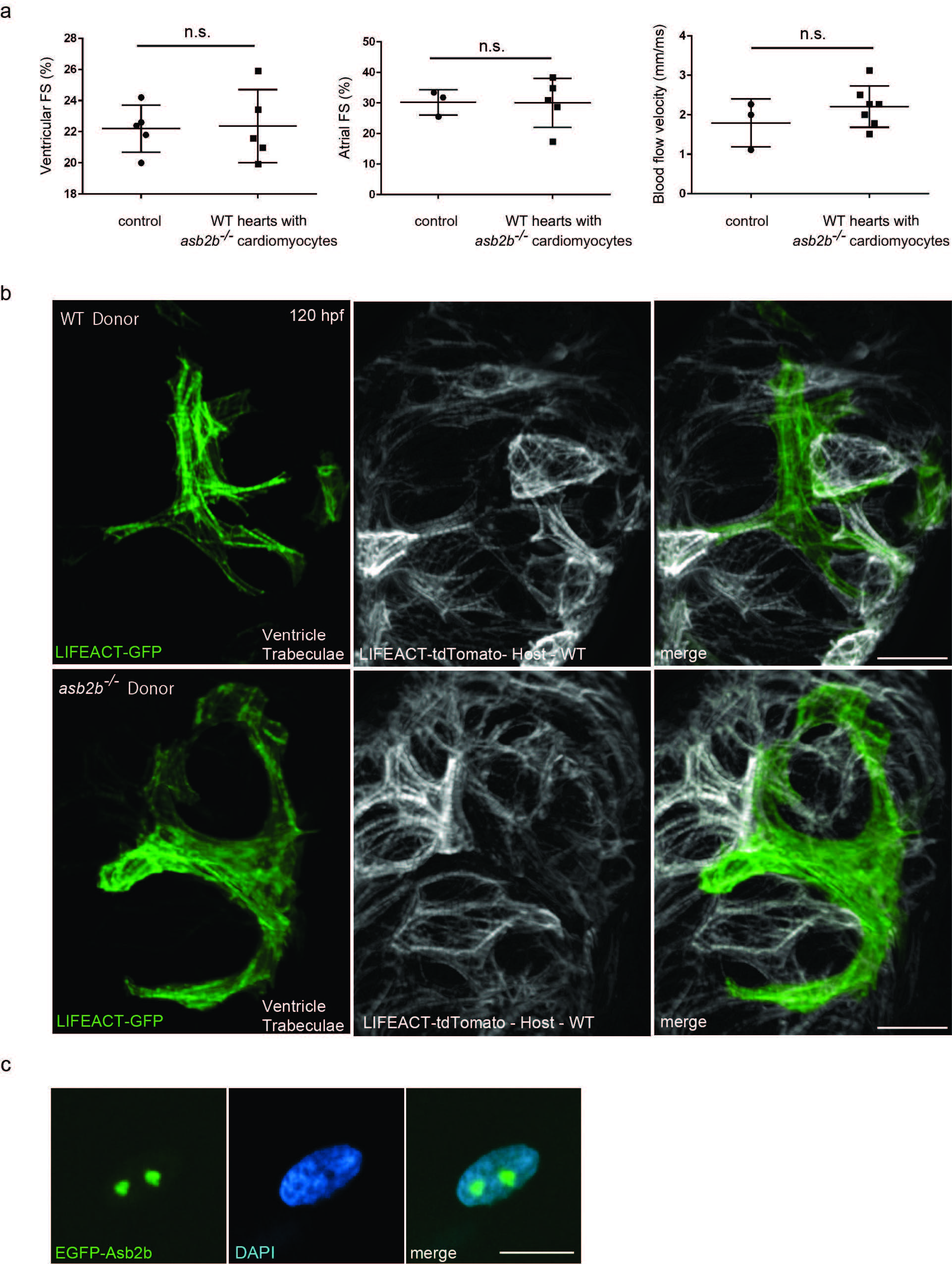
asb2b mutant cardiomyocytes in WT hearts exhibit disorganized myofilaments. (a) Quantification of fractional shortening (FS) in ventricles and atria in 50 hpf chimeric hearts, and aortic blood flow velocity. WT or asb2b mutant cells were transplanted into WT host embryos. No significant differences in FS or blood flow velocity were observed (n=3 to 7 fish). (b) 3D images of 120 hpf chimeric ventricles. Tg(myl7:LIFEACT-GFP) WT or asb2b mutant donor cells were transplanted into Tg(myl7:LIFEACT-tdTomato) WT host embryos. WT donor-derived cardiomyocytes exhibit mature sarcomeres in trabecular cardiomyocytes, while asb2b mutant-derived cardiomyocytes exhibit disorganized sarcomeres. (c) A 50 hpf TgBAC(asb2b:GFP-asb2b) heart stained with DAPI (blue). Scale bars, 20 μm.