
Fig. S2
- ID
- ZDB-IMAGE-170907-27
- Publication
- Pfefferli et al., 2017 - The careg element reveals a common regulation of regeneration in the zebrafish myocardium and fin
- All Figures
- Figures for Pfefferli et al., 2017
|
Fig. S2
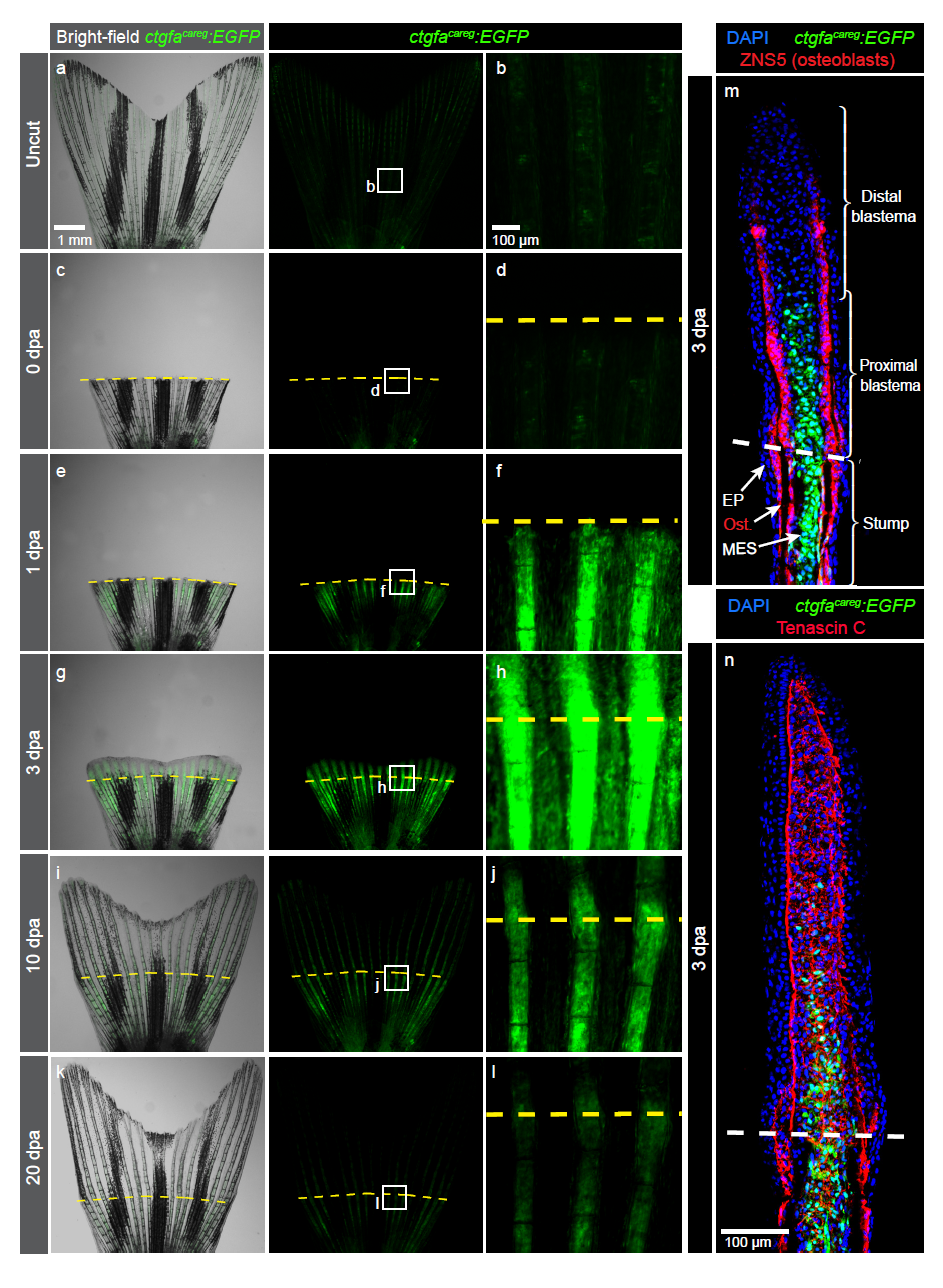
Peri-injury tissue of the fin stump transiently induces ctgfacareg:EGFP expression
(a-l) Live imaging of ctgfacareg:EGFP fins at different days post-amputation (dpa). Higher magnifications of framed areas show the region of the amputation plane (yellow dashed line). N=4. (m-n) Immunofluorescence staining of longitudinal ctgfacareg:EGFP fin sections at 3 dpa. EP, epidermis; Ost, osteoblasts; MES, mesenchmyme. (m) ctgfacareg:EGFP is detected in the stump mesenchyme and osteoblasts (red) below the amputation plane (dashed line), and in the proximal blastema. (n) The mesenchyme of the regenerating fin abundantly expresses Tenascin C, a tissue remodelling extracellular protein. N≥4.