
IMAGE
Fig. 7
- ID
- ZDB-IMAGE-170907-16
- Publication
- Pfefferli et al., 2017 - The careg element reveals a common regulation of regeneration in the zebrafish myocardium and fin
- All Figures
- Figures for Pfefferli et al., 2017
Image
|
Figure Caption
Fig. 7
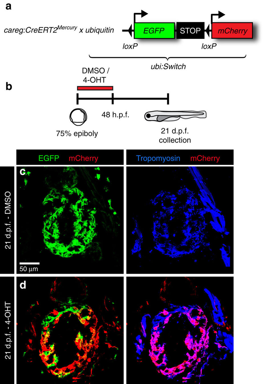
Embryonic careg-positive CMs contribute to the trabecular myocardium.
(a) Schematic representation of the transgenic strains used for lineage tracing. (b) Experimental design. (c,d) Longitudinal sections of hearts at 21 d.p.f. immunostained against mCherry (red), GFP (green) and Tropomyosin (blue). (c) Control embryos treated with the vehicle do not display mCherry fluorescence. (d) mCherry+ CMs are present in the trabecular and outer myocardium at 21 d.p.f. in embryos treated with 4-OHT, suggesting that the trabecular myocardium derives from embryonic careg+ CMs. N=9.
Acknowledgments
This image is the copyrighted work of the attributed author or publisher, and
ZFIN has permission only to display this image to its users.
Additional permissions should be obtained from the applicable author or publisher of the image.
Full text @ Nat. Commun.