
|
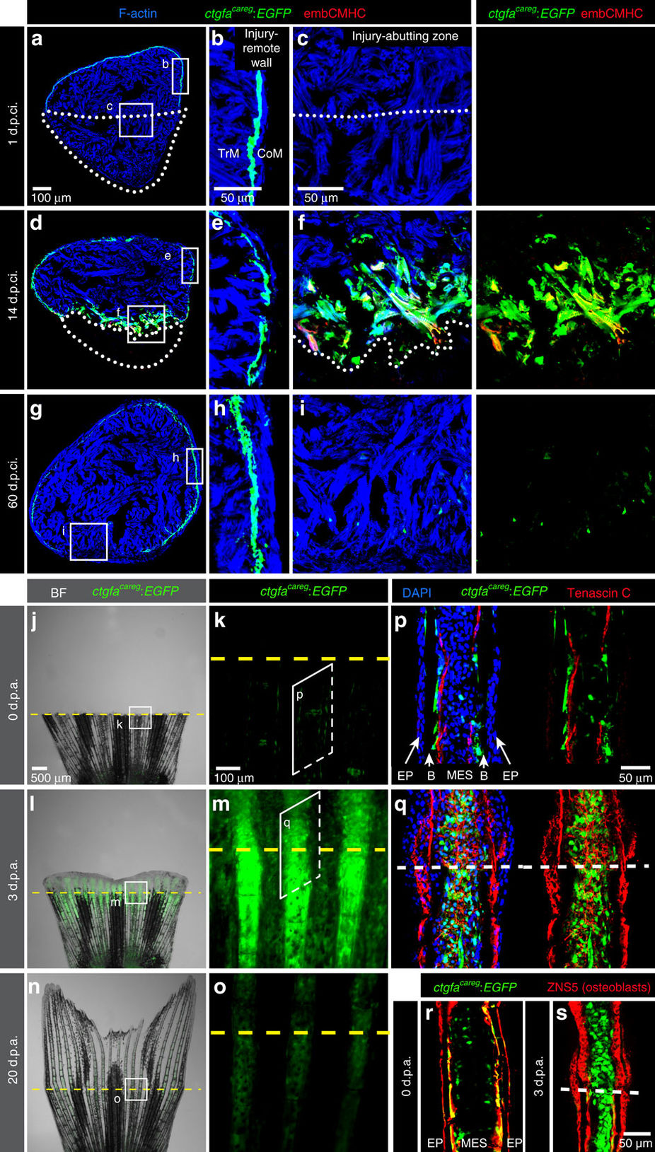
Fig. 1
A transgenic reporter of the peri-injured myocardium and the fin stump.
(a–i) Transversal ventricle sections of transgenic fish ctgfacareg:EGFP immunostained for embCMHC (red) at different days post-cryoinjury (d.p.ci.). The cardiac muscle is detected by F-actin staining (Phalloidin, blue). (b,e,h) The injury-remote part of the ventricular wall displays a subcortical layer of ctgfacareg:EGFP cells that is located between the thin cortical myocardium (CoM) and the inner trabecular myocardium (TrM). This layer does not express embCMHC and remains unaltered during regeneration. (c,f,i) The injury-abutting zone of the ventricular wall shows transient expression of ctgfacareg:EGFP and embCMHC within a distance of 100 μm from the wound border during regeneration. N≥4. (j–o) Live-imaging of fins at different days post amputation (d.p.a.). Bright-field (BF) was combined with fluorescence. (k,m,o) Higher magnifications of the region at the amputation plane (yellow dashed line) show transient expression of ctgfacareg:EGFP in the stump and the regenerate. N=4. (p–s) Immunofluorescence staining of longitudinal fin sections. (p,r) At 0 d.p.a., ctgfacareg:EGFP+ cells are associated with Tenascin C fibres along the border between the bones (B) and mesenchymal cells (MES), but not the epidermis (EP). Some of these cells express an osteoblast marker visualized with Zns5 antibody (red). (q,s) At 3 d.p.a., Tenascin C demarcates the zone of tissue remodelling in the stump and the outgrowth. ctgfacareg:EGFP is upregulated in the activated MES at the peri-injury zone, but not in Zns5-labelled osteoblasts of the outgrowth. N≥4. Post-infarcted ventricle is encircled with a dotted line. Fin amputation plane is shown with a dashed line. The same rules apply to all subsequent figures.