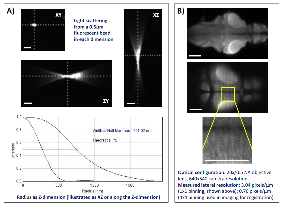
Fig. S3
Summary of achievable 3D and spatial resolution using the LSM system set up used for whole brain neuropharmacological profiling. Panel A) shows the Point Spread Function (PSF) estimation averaged across 50 x 0.5μm fluorescent beads (Emission wavelength: 525nm, Merck Milipore, Haarlerbergweg, The Netherlands) in a 1.5μm step scan using MOSAICsuite (http://imagej.net/MOSAICsuite). The scale bar represents circa. 10μm. Panel B) shows a diagrammatic representation of the spatial resolution achievable with the LSM settings used in the current study. Shown are a maximum intensity projection of the whole scan area (top); resolution in a single slice at the top of this stack (middle); and detail from this slice (bottom) in which individual cell bodies are clearly visible. The scale bar represents circa. 100μm. Images were obtained on the 5.5MP sCMOS camera used throughout the study (30FPS, 640x540 pixels, 4x4 binning, 40ms exposure).

